Luo Jichang, Zhang Xiao, Li Wenjing, Wang Tao, Cui Shengyan, Li Tianhua, Wang Yilin, Xu Wenlong, Ma Yan, Yang Bin, Luo Yumin, Yang Ge, Xu Ran, Jiao Liqun
Department of Neurosurgery, Xuanwu Hospital, Capital Medical University, Beijing, China.
China International Neuroscience Institute (China-INI), Beijing, China.
Heliyon. 2024 Feb 23;10(5):e26904. doi: 10.1016/j.heliyon.2024.e26904. eCollection 2024 Mar 15.
Carotid arterial atherosclerotic stenosis is a well-recognized pathological basis of ischemic stroke; however, its underlying molecular mechanisms remain unknown. Vascular smooth muscle cells (VSMCs) play fundamental roles in the initiation and progression of atherosclerosis. Organelle dynamics have been reported to affect atherosclerosis development. However, the association between organelle dynamics and various cellular stresses in atherosclerotic progression remain ambiguous.
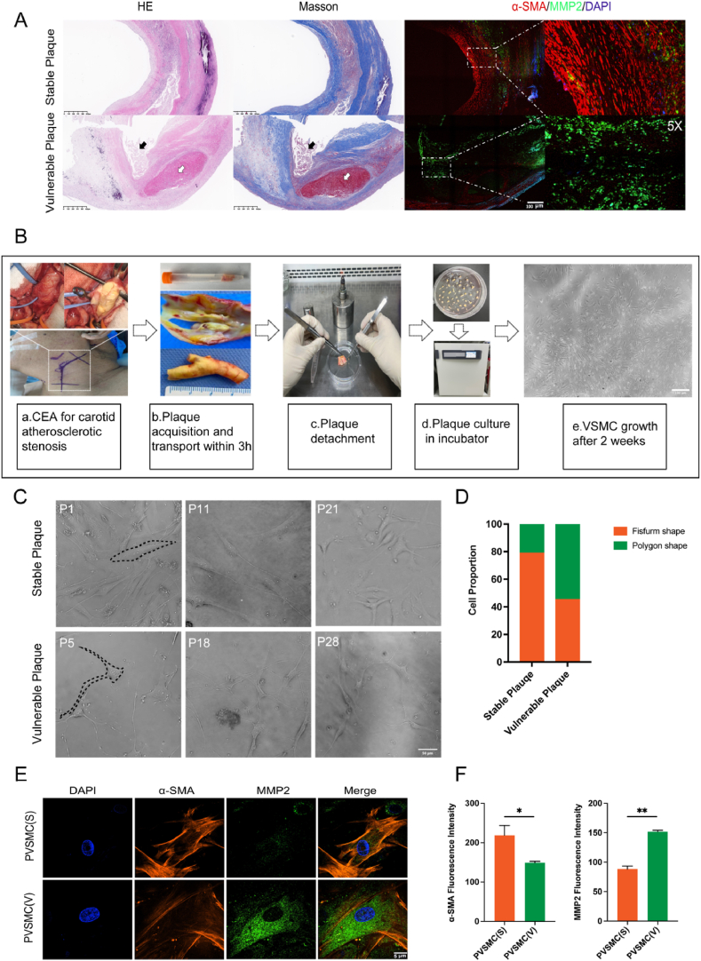
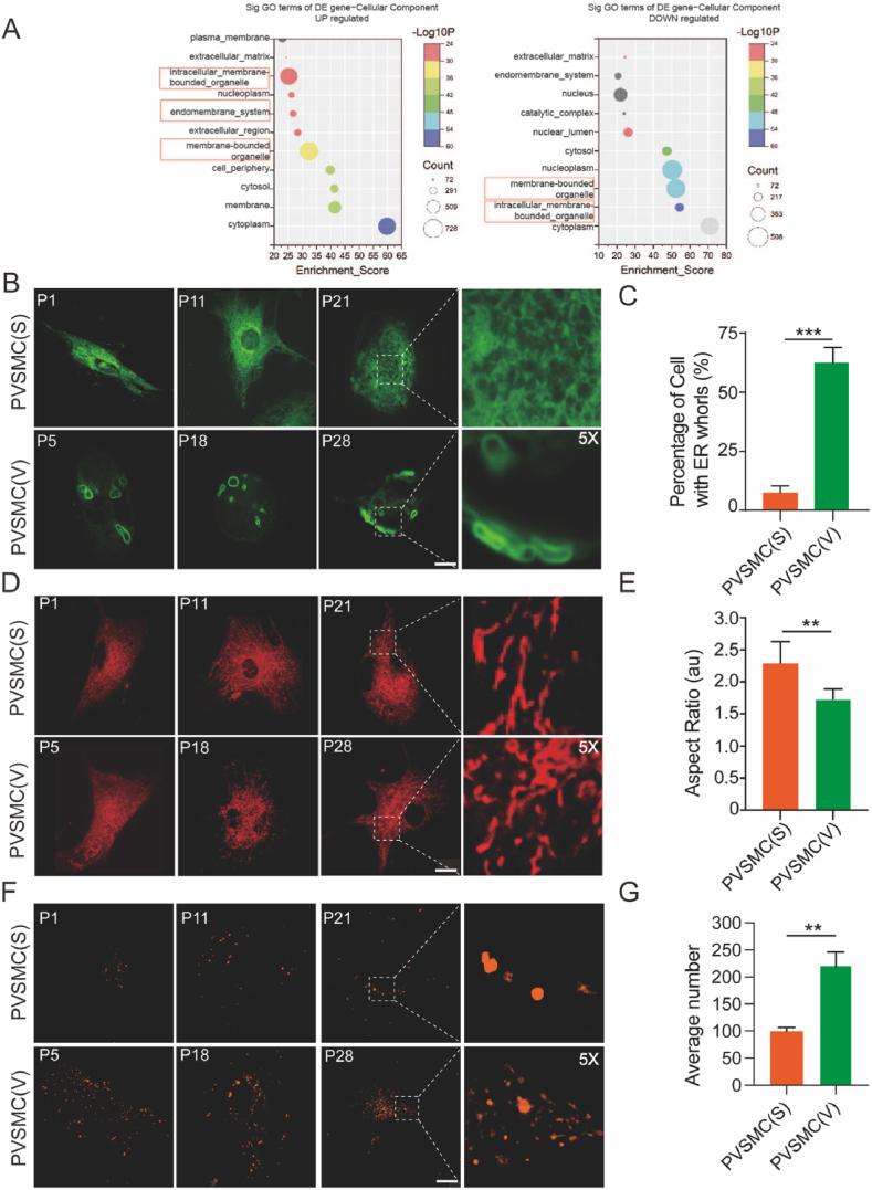
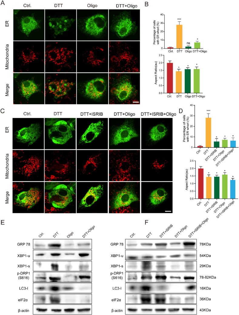
In this study, we conducted transcriptomics and bioinformatics analyses of stable and vulnerable carotid plaques. Primary VSMCs were isolated from carotid plaques and subjected to histopathological staining to determine their expression profiles. Endoplasmic reticulum (ER), mitochondria, and lysosome dynamics were observed in primary VSMCs and VSMC cell lines using live-cell imaging. Moreover, the mechanisms underlying disordered organelle dynamics were investigated using comprehensive biological approaches.
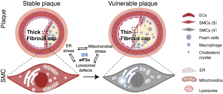
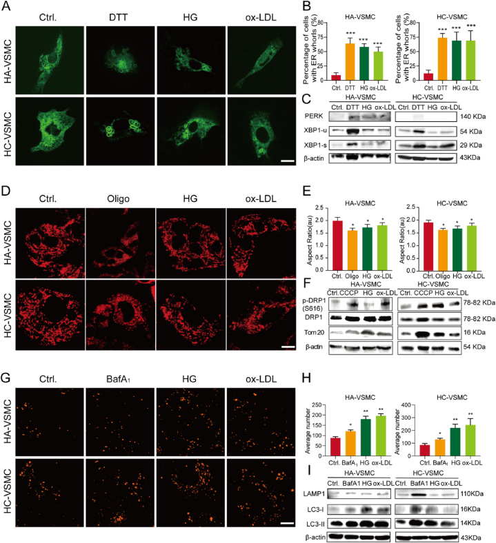
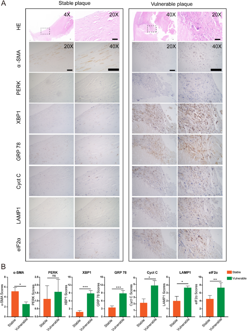
ER whorls, a representative structural change under ER stress, are prominent dynamic reconstructions of VSMCs between vulnerable and stable plaques, followed by fragmented mitochondria and enlarged lysosomes, suggesting mitochondrial stress and lysosomal defects, respectively. Induction of mitochondrial stress alleviated ER stress and autophagy in an eukaryotic translation initiation factor (eIF)-2α-dependent manner. Furthermore, the effects of eIF2α on ER stress, mitochondrial stress, and lysosomal defects were validated using clinical samples.
Our results indicate that morphological and functional changes in VSMC organelles, especially in ER whorls, can be used as reliable biomarkers for atherosclerotic progression. Moreover, eIF2α plays an important role in integrating multiple stress-signaling pathways to determine the behavior and fate of VSMCs.
颈动脉粥样硬化狭窄是公认的缺血性中风的病理基础;然而,其潜在的分子机制仍不清楚。血管平滑肌细胞(VSMC)在动脉粥样硬化的发生和发展中起重要作用。据报道,细胞器动态变化会影响动脉粥样硬化的发展。然而,在动脉粥样硬化进展过程中,细胞器动态变化与各种细胞应激之间的关联仍不明确。
在本研究中,我们对稳定和易损颈动脉斑块进行了转录组学和生物信息学分析。从颈动脉斑块中分离出原代VSMC,并进行组织病理学染色以确定其表达谱。使用活细胞成像技术观察原代VSMC和VSMC细胞系中的内质网(ER)、线粒体和溶酶体动态变化。此外,采用综合生物学方法研究细胞器动态紊乱的潜在机制。
内质网应激下的典型结构变化——内质网涡,是易损斑块和稳定斑块之间VSMC的显著动态重构,其次是线粒体碎片化和溶酶体增大,分别提示线粒体应激和溶酶体缺陷。线粒体应激的诱导以真核翻译起始因子(eIF)-2α依赖的方式减轻内质网应激和自噬。此外,使用临床样本验证了eIF2α对内质网应激、线粒体应激和溶酶体缺陷的影响。
我们的结果表明,VSMC细胞器的形态和功能变化,尤其是内质网涡,可作为动脉粥样硬化进展的可靠生物标志物。此外,eIF2α在整合多种应激信号通路以决定VSMC的行为和命运方面起重要作用。