Department of Ophthalmology, Faculty of Medicine, Universiti Kebangsaan Malaysia (UKM) Medical Centre, Sakaka, Al-Jawf Province, Saudi Arabia.
Department of Clinical Laboratory Sciences, College of Applied Medical Sciences, Jouf University, Sakaka, Al-Jawf Province, Saudi Arabia; Department of Biomedical Science, Faculty of Medicine and Health Sciences, Universiti Putra Malaysia, Serdang, Malaysia.
Indian J Ophthalmol. 2022 Mar;70(3):921-929. doi: 10.4103/ijo.IJO_472_21.
This study aimed to investigate the efficacy of human-derived umbilical cord mesenchymal stem cells (HDUMSC) and human-derived umbilical cord mesenchymal stem cells expressing erythropoietin (HDUMSC-EPO) to rescue total degenerated retina in a rat model.
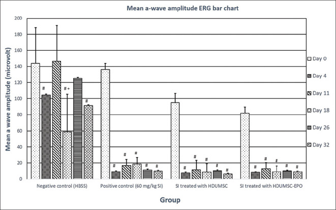
The study included four treatment groups, namely negative control using normal saline (HBSS) injection, positive control using sodium iodide 60 mg/kg (SI), SI treated with HDUMSC, and SI treated with HDUMSC-EPO given via subretinal and intravenous routes, to test the efficacy of retinal regeneration following SI-induced retinal degeneration. Retinal function in both phases was tested via electroretinography (ERG) and histological staining examining the outer nuclear layer (ONL).
There was a statistically significant result (P < 0.05) in the SI treated with HDUMSC-EPO only when comparing day 11 (mean = 23.6 μv), day 18 (mean = 25.2 μv), day 26 (mean = 26.3 μv), and day 32 (mean = 28.2 μv) to the b-wave ERG on day 4 rescue injection day (mean = 12.5 μv). The SI treated with HDUMSC-EPO showed significant improvement in b-wave ERG readings in the Sprague-Dawley (SD) rat but did not restore baseline readings prior to degeneration (day 0). Both treated groups' ONL thicknesses did not show significant changes compared to the negative control group (HBSS) following rescue therapy.
Total retinal degeneration following intravenous SI injection was observed at 60 mg/kg. SI treated with HDUMSC and HDUMSC-EPO showed no regenerative potential compared to baseline in SI-induced total retina degeneration on ERG or histology, whereas SI treated with HDUMSC-EPO group showed a substantial increase in b-wave ERG amplitude over time.
本研究旨在探讨人源性脐带间充质干细胞(HDUMSC)和表达促红细胞生成素的人源性脐带间充质干细胞(HDUMSC-EPO)对大鼠全退化视网膜的治疗效果。
本研究包括四个治疗组,即生理盐水(HBSS)注射阴性对照组、60mg/kg 碘酸钠(SI)阳性对照组、SI 联合 HDUMSC 治疗组和 SI 联合 HDUMSC-EPO 经视网膜下和静脉途径治疗组,以测试 SI 诱导的视网膜变性后视网膜再生的效果。通过视网膜电图(ERG)和外核层(ONL)组织学染色检测评估视网膜功能在两个阶段的变化。
仅在 SI 联合 HDUMSC-EPO 治疗组中观察到统计学上的显著差异(P<0.05),在第 11 天(平均值=23.6μv)、第 18 天(平均值=25.2μv)、第 26 天(平均值=26.3μv)和第 32 天(平均值=28.2μv)与第 4 天挽救性注射日(平均值=12.5μv)的 b 波 ERG 进行比较时。SI 联合 HDUMSC-EPO 治疗组在 Sprague-Dawley(SD)大鼠的 b 波 ERG 读数上显示出显著改善,但在退行性病变前(第 0 天)未恢复基线读数。与阴性对照组(HBSS)相比,在挽救治疗后,两组治疗组的 ONL 厚度均无明显变化。
静脉注射 60mg/kg SI 后可观察到全视网膜变性。与 SI 诱导的全视网膜变性的 ERG 或组织学基线相比,SI 联合 HDUMSC 和 HDUMSC-EPO 治疗组均未显示出再生潜力,而 SI 联合 HDUMSC-EPO 组的 b 波 ERG 幅度随时间显著增加。