Mandai Michiko, Fujii Momo, Hashiguchi Tomoyo, Sunagawa Genshiro A, Ito Shin-ichiro, Sun Jianan, Kaneko Jun, Sho Junki, Yamada Chikako, Takahashi Masayo
Laboratory for Retinal Regeneration, RIKEN Center for Developmental Biology, 2-2-3, Minatojima-minamimachi, Chuo-ku, Kobe, Hyogo 650-0047, Japan.
Laboratory for Retinal Regeneration, RIKEN Center for Developmental Biology, 2-2-3, Minatojima-minamimachi, Chuo-ku, Kobe, Hyogo 650-0047, Japan.
Stem Cell Reports. 2017 Jan 10;8(1):69-83. doi: 10.1016/j.stemcr.2016.12.008.
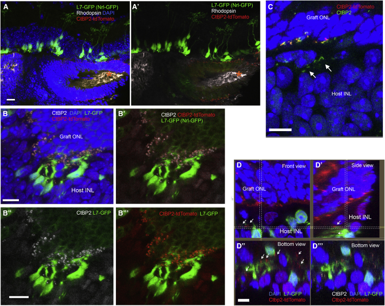
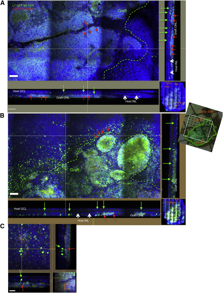
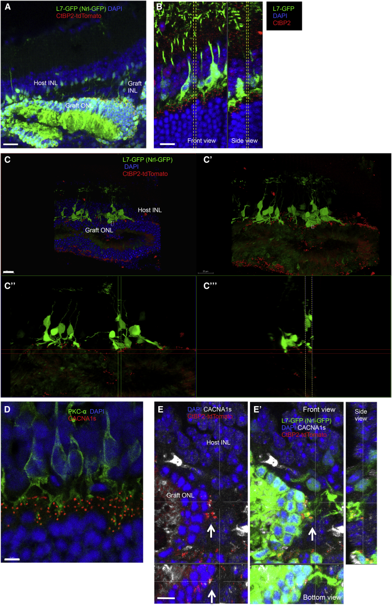
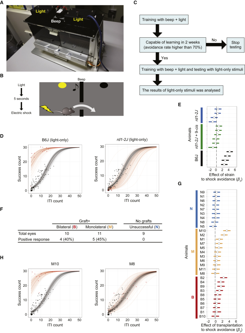
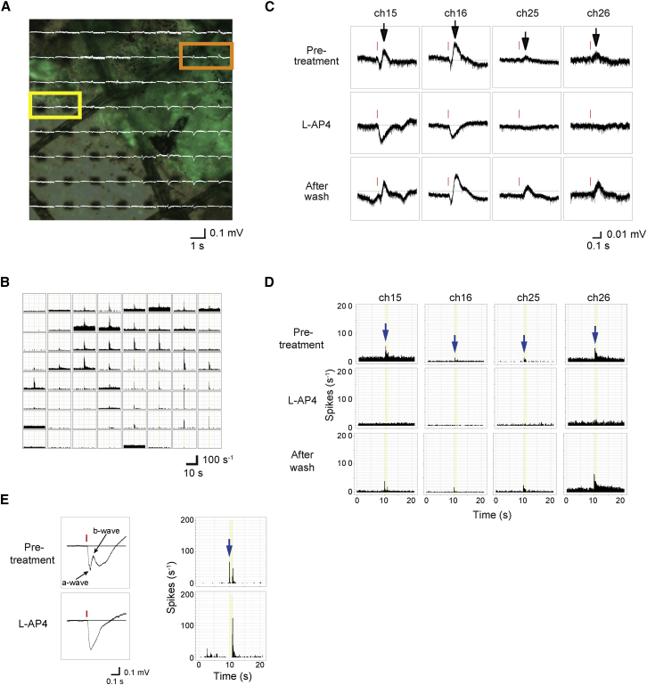
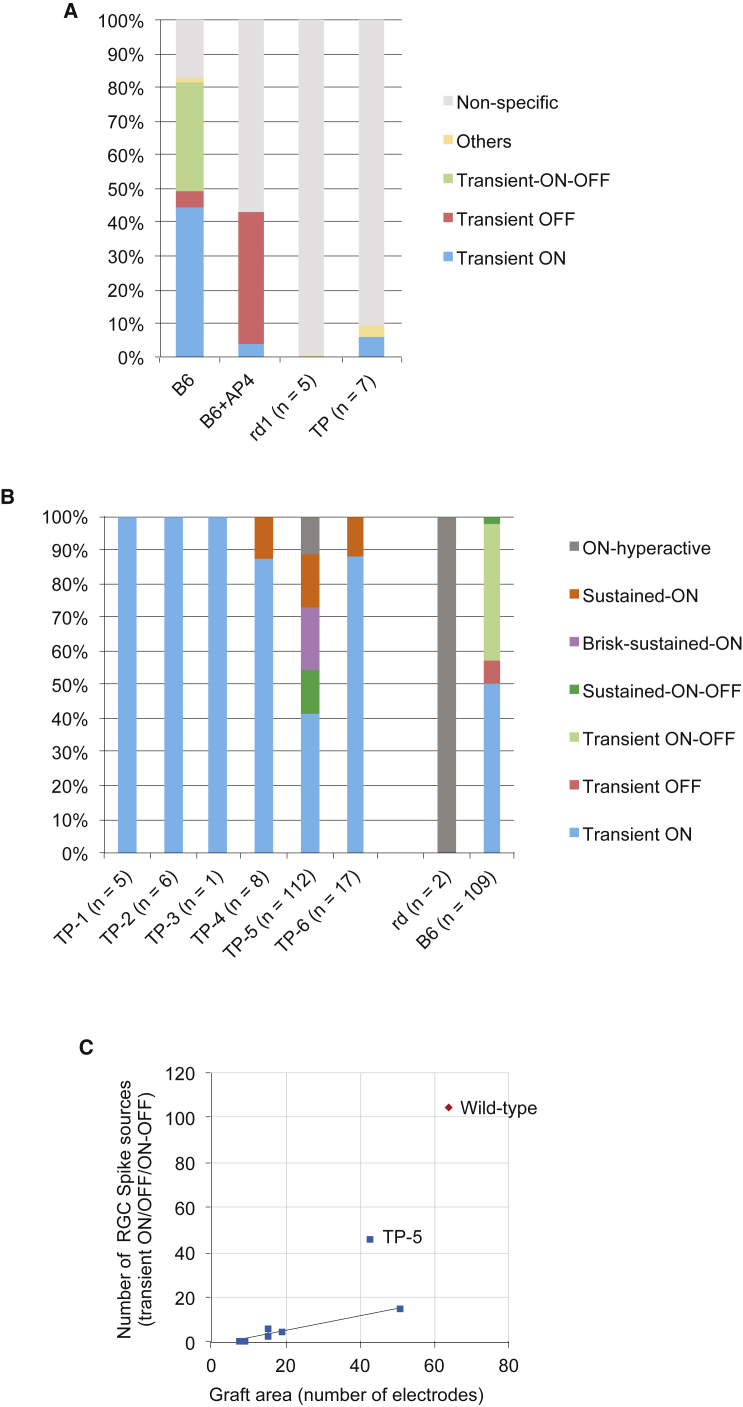
Recent success in functional recovery by photoreceptor precursor transplantation in dysfunctional retina has led to an increased interest in using embryonic stem cell (ESC) or induced pluripotent stem cell (iPSC)-derived retinal progenitors to treat retinal degeneration. However, cell-based therapies for end-stage degenerative retinas that have lost the outer nuclear layer (ONL) are still a big challenge. In the present study, by transplanting mouse iPSC-derived retinal tissue (miPSC retina) in the end-stage retinal-degeneration model (rd1), we visualized the direct contact between host bipolar cell terminals and the presynaptic terminal of graft photoreceptors by gene labeling, showed light-responsive behaviors in transplanted rd1 mice, and recorded responses from the host retina with transplants by ex vivo micro-electroretinography and ganglion cell recordings using a multiple-electrode array system. Our data provides a proof of concept for transplanting ESC/iPSC retinas to restore vision in end-stage retinal degeneration.
最近,通过将光感受器前体细胞移植到功能失调的视网膜中实现功能恢复取得了成功,这使得人们对使用胚胎干细胞(ESC)或诱导多能干细胞(iPSC)衍生的视网膜祖细胞来治疗视网膜变性的兴趣日益增加。然而,对于已经失去外核层(ONL)的终末期退行性视网膜,基于细胞的治疗仍然是一个巨大的挑战。在本研究中,我们将小鼠iPSC衍生的视网膜组织(miPSC视网膜)移植到终末期视网膜变性模型(rd1)中,通过基因标记观察到宿主双极细胞终末与移植的光感受器突触前终末之间的直接接触,在移植的rd1小鼠中显示出光反应行为,并使用多电极阵列系统通过离体微视网膜电图和神经节细胞记录来记录宿主视网膜与移植组织的反应。我们的数据为移植ESC/iPSC视网膜以恢复终末期视网膜变性患者的视力提供了概念验证。