Chen Qi, Chu Ling, Li Xinyu, Li Hao, Zhang Ying, Cao Qingtai, Zhuang Quan
Transplantation Center, Third Xiangya Hospital, Central South University, Changsha, China.
Xiangya School of Medicine, Central South University, Changsha, China.
Front Cell Dev Biol. 2022 Feb 14;9:801715. doi: 10.3389/fcell.2021.801715. eCollection 2021.
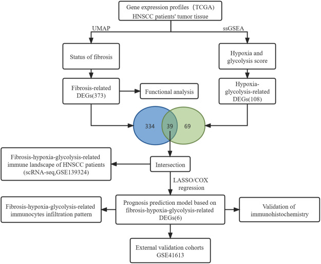
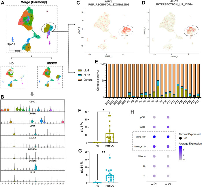
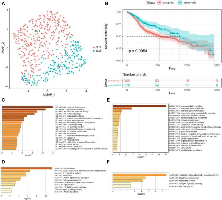
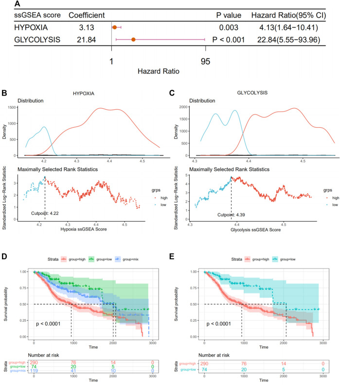
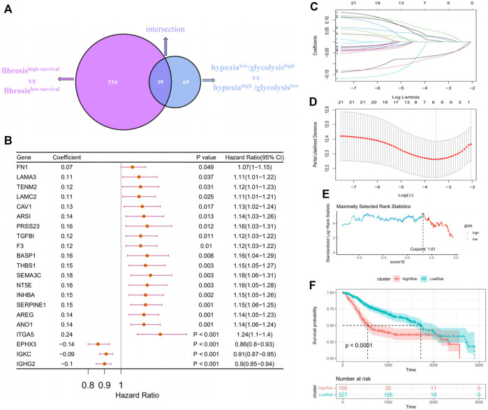
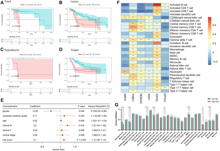
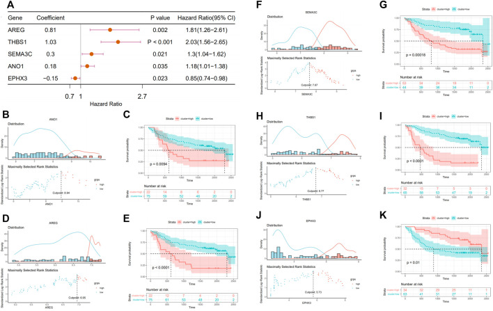
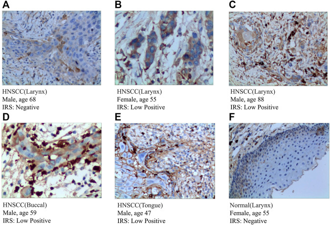
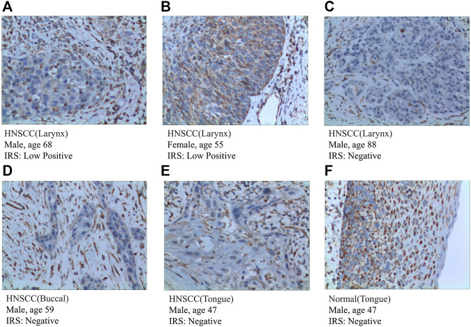
There is accumulating evidence on the clinical importance of the fibroblast growth factor receptor (FGFR) signal, hypoxia, and glycolysis in the immune microenvironment of head and neck squamous cell carcinoma (HNSCC), yet reliable prognostic signatures based on the combination of the fibrosis signal, hypoxia, and glycolysis have not been systematically investigated. Herein, we are committed to establish a fibrosis-hypoxia-glycolysis-related prediction model for the prognosis and related immune infiltration of HNSCC. Fibrotic signal status was estimated with microarray data of a discovery cohort from the TCGA database using the UMAP algorithm. Hypoxia, glycolysis, and immune-cell infiltration scores were imputed using the ssGSEA algorithm. Cox regression with the LASSO method was applied to define prognostic genes and develop a fibrosis-hypoxia-glycolysis-related gene signature. Immunohistochemistry (IHC) was conducted to identify the expression of specific genes in the prognostic model. Protein expression of several signature genes was evaluated in HPA. An independent cohort from the GEO database was used for external validation. Another scRNA-seq data set was used to clarify the related immune infiltration of HNSCC. Six genes, including AREG, THBS1, SEMA3C, ANO1, IGHG2, and EPHX3, were identified to construct a prognostic model for risk stratification, which was mostly validated in the independent cohort. Multivariate analysis revealed that risk score calculated by our prognostic model was identified as an independent adverse prognostic factor ( < .001). Activated B cells, immature B cells, activated CD4 T cells, activated CD8 T cells, effector memory CD8 T cells, MDSCs, and mast cells were identified as key immune cells between high- and low-risk groups. IHC results showed that the expression of SEMA3C, IGHG2 were slightly higher in HNSCC tissue than normal head and neck squamous cell tissue. THBS1, ANO1, and EPHX3 were verified by IHC in HPA. By using single-cell analysis, FGFR-related genes and highly expressed DEGs in low-survival patients were more active in monocytes than in other immune cells. A fibrosis-hypoxia-glycolysis-related prediction model provides risk estimation for better prognoses to patients diagnosed with HNSCC.
关于成纤维细胞生长因子受体(FGFR)信号、缺氧和糖酵解在头颈部鳞状细胞癌(HNSCC)免疫微环境中的临床重要性,已有越来越多的证据,但基于纤维化信号、缺氧和糖酵解组合的可靠预后特征尚未得到系统研究。在此,我们致力于建立一个与HNSCC预后及相关免疫浸润相关的纤维化-缺氧-糖酵解预测模型。使用UMAP算法,通过TCGA数据库中一个发现队列的微阵列数据评估纤维化信号状态。使用ssGSEA算法估算缺氧、糖酵解和免疫细胞浸润评分。应用LASSO方法的Cox回归来定义预后基因并开发一个与纤维化-缺氧-糖酵解相关的基因特征。进行免疫组织化学(IHC)以鉴定预后模型中特定基因的表达。在人类蛋白质图谱(HPA)中评估几个特征基因的蛋白质表达。使用来自GEO数据库的独立队列进行外部验证。另一个单细胞RNA测序(scRNA-seq)数据集用于阐明HNSCC的相关免疫浸润。鉴定出包括AREG、THBS1、SEMA3C、ANO1、IGHG2和EPHX3在内的六个基因,以构建用于风险分层的预后模型,该模型在独立队列中得到了大部分验证。多变量分析显示,由我们的预后模型计算出的风险评分被确定为独立的不良预后因素(<0.001)。活化B细胞、未成熟B细胞、活化CD4 T细胞、活化CD8 T细胞、效应记忆CD8 T细胞、髓系来源的抑制细胞(MDSCs)和肥大细胞被确定为高风险组和低风险组之间的关键免疫细胞。IHC结果显示,SEMA3C、IGHG2在HNSCC组织中的表达略高于正常头颈部鳞状细胞组织。THBS1、ANO1和EPHX3在HPA中通过IHC得到验证。通过单细胞分析,FGFR相关基因和低生存患者中高表达的差异表达基因(DEGs)在单核细胞中比在其他免疫细胞中更活跃。一个与纤维化-缺氧-糖酵解相关的预测模型为诊断为HNSCC的患者提供更好预后的风险估计。