Hofmann Sarah, Cohen-Harazi Raichel, Maizels Yael, Koman Igor
Institute for Personalized and Translational Medicine, Ariel University, Ariel, Israel.
Brigham and Women's Hospital, Boston, MA, USA.
Transl Cancer Res. 2022 Jan;11(1):134-147. doi: 10.21037/tcr-21-1577.
Breast cancer is the most common cause of cancer related deaths in women. Treatment of breast cancer has many limitations including a lack of accurate biomarkers to predict success of chemotherapy and intrinsic resistance of a significant group of patients to the gold standard of therapy. Therefore, new tools are needed to provide doctors with guidance in choosing the most effective treatment plan for a particular patient and thus to increase the survival rate for breast cancer patients.
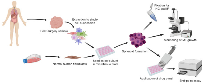
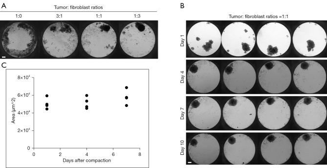
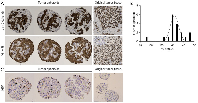
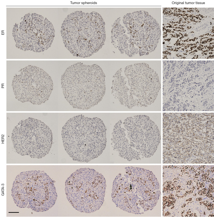
Here, we present a successful method to grow spheroids from primary breast cancer tissue. Samples were received in accordance with relevant ethical guidelines and regulations. After tissue dissociation, spheroids were generated in a scaffold-free 96-well plate format. Spheroid composition was investigated by immunohistochemistry (IHC) of epithelial [pan cytokeratin (panCK)], stromal (vimentin) and breast cancer-specific markers (ER, PR, HER2, GATA). Growth and cell viability of the spheroids were assessed upon treatment with multiple anti-cancer compounds. Student's -test and two-way ANOVA test were used to determine statistical significance.
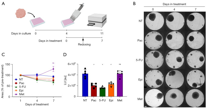
We were able to successfully grow spheroids from 27 out of 31 samples from surgical resections of breast cancer tissue from previously untreated patients. Recapitulation of the histopathology of the tissue of origin was confirmed. Furthermore, a drug panel of standard first-line chemotherapy drugs used to treat breast cancer was applied to assess the viability of the patient-derived spheroids and revealed variation between samples in the response of the spheroids to different drug treatments.
We investigated the feasibility and the utility of an , patient-derived spheroid model for breast cancer therapy, and we conclude that spheroids serve as a highly effective platform to explore cancer therapeutics and personalized treatment efficacy. These results have significant implications for the application of this model in clinical personalized medicine.
乳腺癌是女性癌症相关死亡的最常见原因。乳腺癌治疗存在诸多局限性,包括缺乏准确的生物标志物来预测化疗效果,以及相当一部分患者对标准治疗方案存在内在抗性。因此,需要新的工具来指导医生为特定患者选择最有效的治疗方案,从而提高乳腺癌患者的生存率。
在此,我们展示了一种从原发性乳腺癌组织培养球体的成功方法。样本的获取符合相关伦理准则和规定。组织解离后,在无支架的96孔板中生成球体。通过上皮细胞(全细胞角蛋白(panCK))、基质细胞(波形蛋白)和乳腺癌特异性标志物(雌激素受体(ER)、孕激素受体(PR)、人表皮生长因子受体2(HER2)、GATA)的免疫组织化学(IHC)研究球体组成。在用多种抗癌化合物处理后评估球体的生长和细胞活力。使用学生t检验和双向方差分析来确定统计学意义。
我们成功地从31例未经治疗患者的乳腺癌手术切除组织样本中的27例培养出球体。证实了原发组织组织病理学的重现性。此外,应用用于治疗乳腺癌的标准一线化疗药物组成的药物组合来评估患者来源球体的活力,并揭示了不同样本中球体对不同药物治疗反应的差异。
我们研究了一种患者来源的乳腺癌球体模型的可行性和实用性,我们得出结论,球体是探索癌症治疗和个性化治疗效果的高效平台。这些结果对该模型在临床个性化医学中的应用具有重要意义。