Huang Daniel, Watal Pankaj, Drehner Dennis, Dhar Deeksha, Chandra Tushar
Radiology, University of Central Florida College of Medicine, Orlando, USA.
Radiology, Nemours Childrens Hospital, Orlando, USA.
Cureus. 2022 Feb 3;14(2):e21863. doi: 10.7759/cureus.21863. eCollection 2022 Feb.
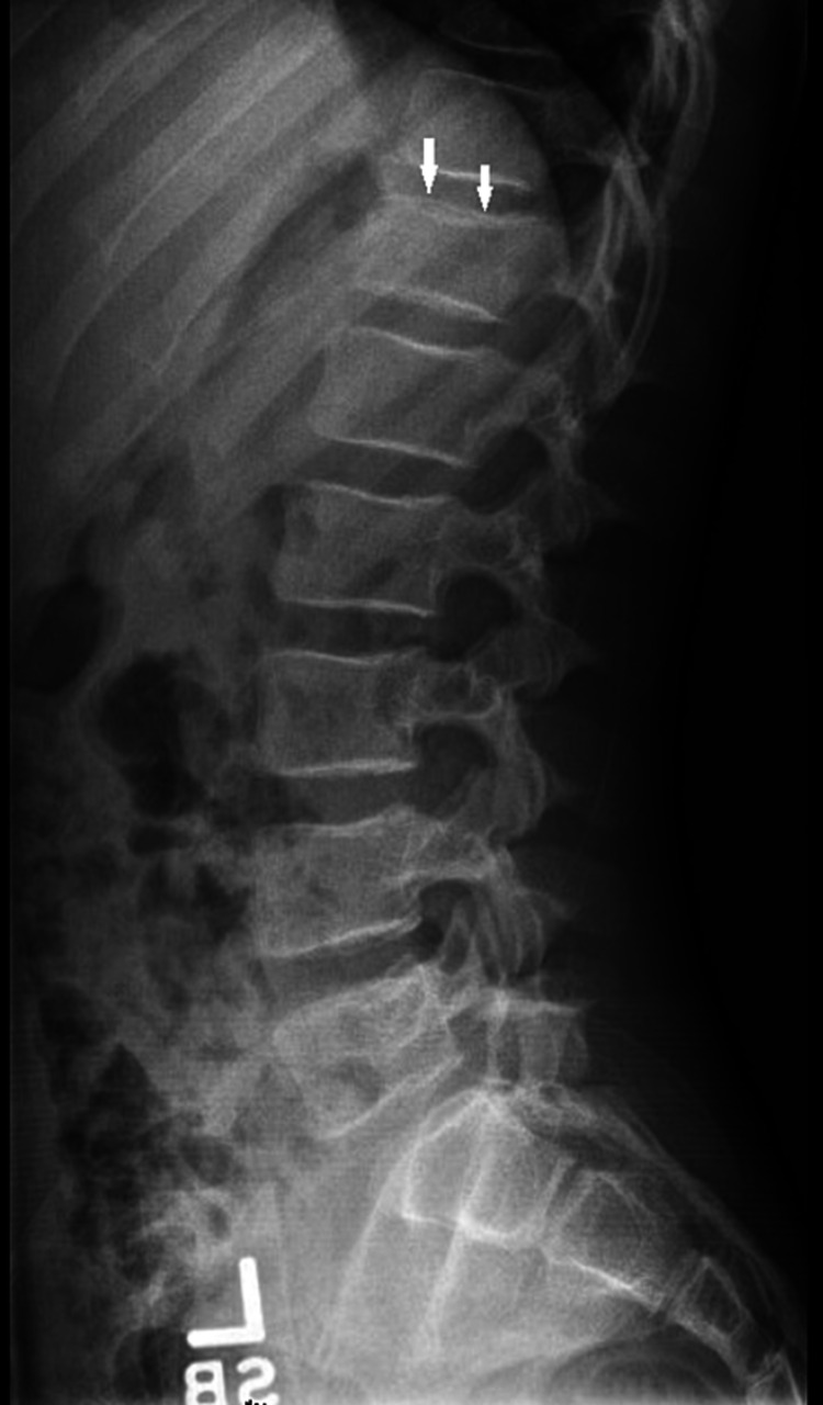
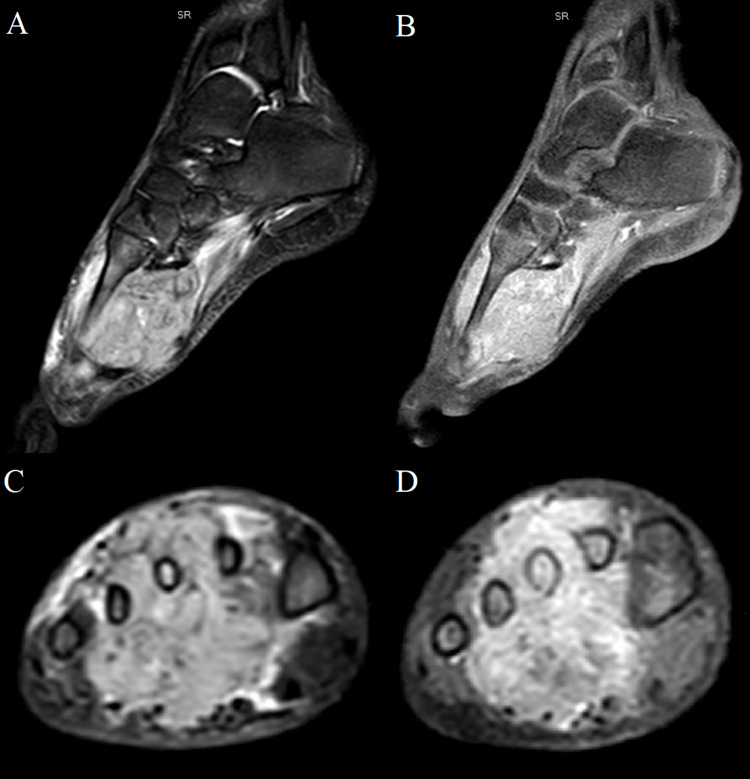
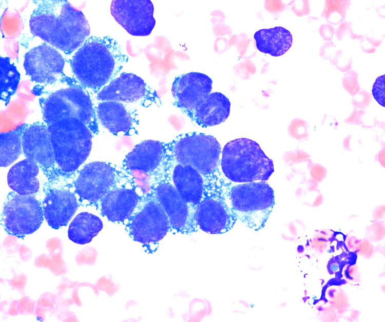
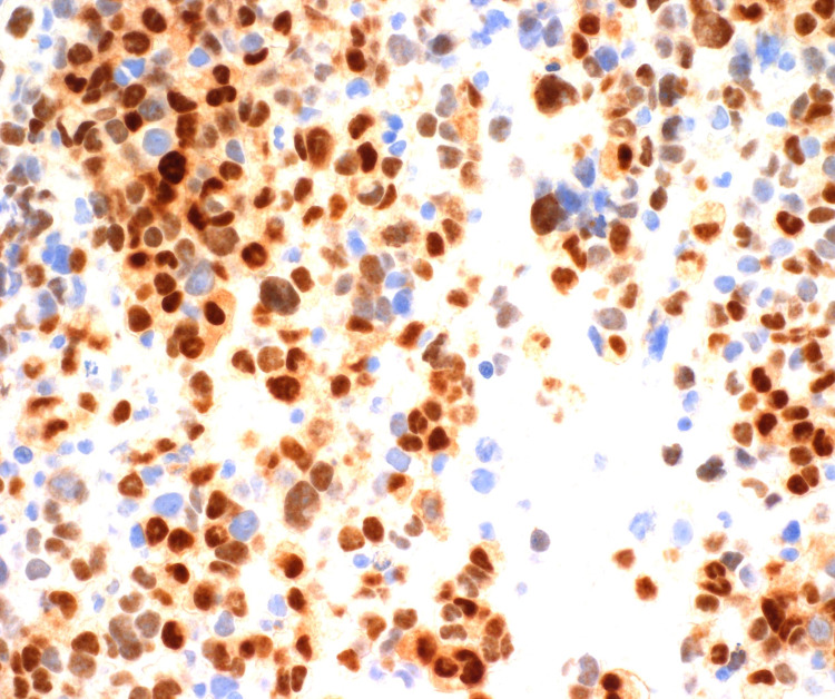
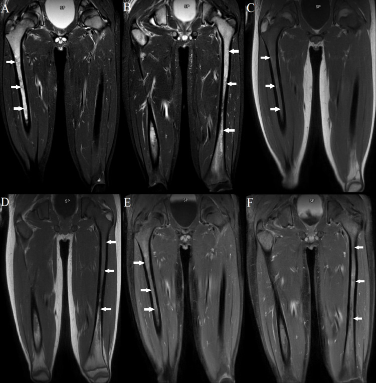
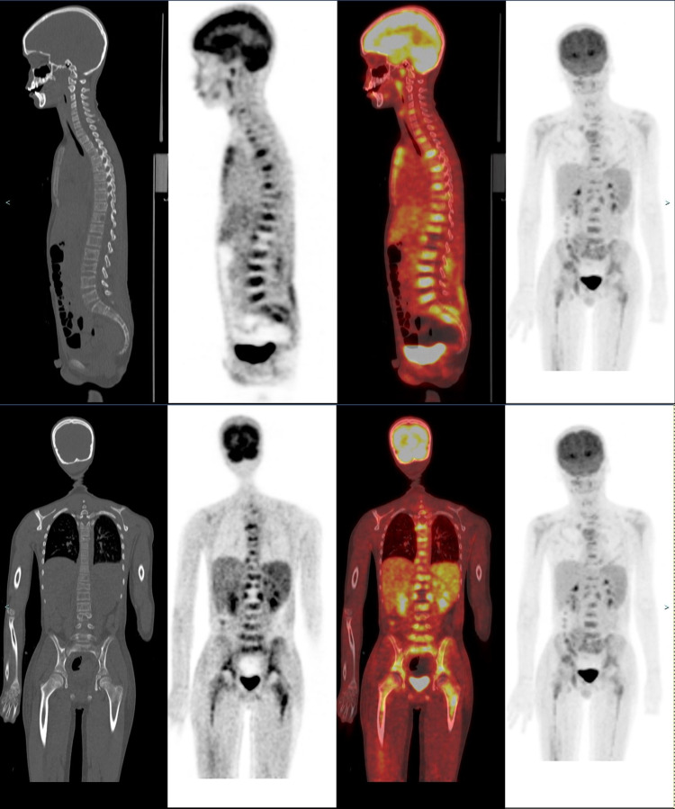
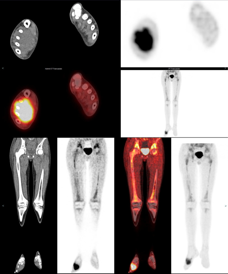
Rhabdomyosarcoma is a highly aggressive cancer that is generally considered a disease of childhood. A vast majority of cases occur in those below the age of 20. Rhabdomyosarcoma can occur in any soft tissue in the body but is primarily found in the head, neck, orbit, genitourinary tract, genitals, and extremities. Prognosis is closely tied to the location of the primary tumor and the extent of metastatic spread. As with most sarcomas, rhabdomyosarcoma has a pattern of hematogenous spread which favors metastasis to the lungs. Other common areas include bone marrows, liver, breasts, and brain. One unusual pattern is the presence of diffuse bone marrow metastases in absence of significant soft tissue disease other than primary (no distant nodal disease, absence of visceral disease in chest and abdomen). Frequently in such cases, patients may have initial presentation similar to hematologic malignancy especially when the primary tumor is not evident. This pattern has been rarely described in the radiology literature. This pattern appears to be well documented in pathology literature. Even more rarely, in some cases, the primary tumor site may not be found after imaging and may remain undetermined even postmortem - only diagnosed by bone marrow aspiration. Awareness of this unique pattern is clearly important for radiologists, especially pediatric radiologists, as misdiagnosis can lead to delay in appropriate treatment that ultimately results in increased mortality. We present a case of rhabdomyosarcoma with this unique pattern of bone marrow metastases in which initial differential diagnosis favored a leukemic picture. This paper will go over the diagnostic techniques utilized throughout our patient's disease course as well as treatment.
横纹肌肉瘤是一种侵袭性很强的癌症,通常被认为是一种儿童疾病。绝大多数病例发生在20岁以下的人群中。横纹肌肉瘤可发生于身体的任何软组织,但主要见于头部、颈部、眼眶、泌尿生殖道、生殖器和四肢。预后与原发肿瘤的位置和转移扩散的程度密切相关。与大多数肉瘤一样,横纹肌肉瘤有血行转移的模式,容易转移到肺部。其他常见部位包括骨髓、肝脏、乳房和大脑。一种不寻常的模式是在除原发灶外无明显软组织疾病(无远处淋巴结疾病,胸部和腹部无内脏疾病)的情况下出现弥漫性骨髓转移。在这种情况下,患者通常最初的表现类似于血液系统恶性肿瘤,尤其是在原发肿瘤不明显时。这种模式在放射学文献中很少被描述。这种模式在病理学文献中似乎有充分的记录。更罕见的是,在某些情况下,影像学检查后可能找不到原发肿瘤部位,甚至死后仍无法确定——只能通过骨髓穿刺诊断。认识到这种独特的模式对放射科医生,尤其是儿科放射科医生显然很重要,因为误诊可能导致适当治疗的延迟,最终导致死亡率增加。我们报告一例具有这种独特骨髓转移模式的横纹肌肉瘤病例,其最初的鉴别诊断倾向于白血病表现。本文将介绍在我们患者的疾病过程中所采用的诊断技术以及治疗方法。