Hugon Gaëlle, Goutal Sébastien, Sarazin Marie, Bottlaender Michel, Caillé Fabien, Droguerre Marine, Charvériat Mathieu, Winkeler Alexandra, Tournier Nicolas
Université Paris-Saclay, Inserm, CNRS, CEA, Laboratoire d'Imagerie Biomédicale Multimodale (BioMaps), Service Hospitalier Frédéric Joliot, Orsay, France.
Department of Neurology of Memory and Language, GHU Paris Psychiatry and Neurosciences, Paris, France.
Front Neurosci. 2022 Feb 25;16:835577. doi: 10.3389/fnins.2022.835577. eCollection 2022.
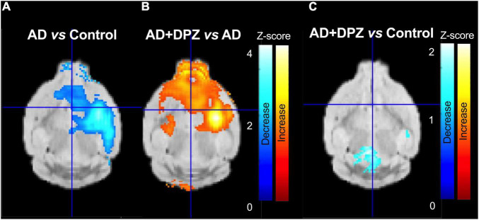
Translational methods are needed to monitor the impact of the Alzheimer's disease (AD) and therapies on brain function in animal models and patients. The formation of amyloid plaques was investigated using [F]florbetapir autoradiography in a mouse model of AD consisting in unilateral intracerebroventricular (i.c.v) injection of amyloid peptide Aβ. Then, an optimized positron emission tomography (PET) imaging protocol using [F]2-fluoro-2-deoxy-D-glucose ([F]FDG) was performed to estimate brain glucose metabolism: [F]FDG was injected in awake animals to allow for 40 min brain uptake in freely moving mice. Anesthesia was then induced for 30 min PET acquisition to capture the slow and poorly reversible brain uptake of [F]FDG. Impact of donepezil (0.25 mg/kg daily, 7 days, orally) on brain function was investigated in AD mice ( = 6 mice/group). Formation of amyloid plaques could not be detected using autoradiography. Compared with sham controls (injection of scramble peptide), significant decrease in [F]FDG uptake was observed in the AD group in the subcortical volume of the ipsilateral hemisphere. Donepezil restored normal glucose metabolism by selectively increasing glucose metabolism in the affected subcortical volume but not in other brain regions. In mice, [F]FDG PET imaging can be optimized to monitor impaired brain function associated with i.c.v injection of Aβ, even in the absence of detectable amyloid plaque. This model recapitulates the regional decrease in [F]FDG uptake observed in AD patients. [F]FDG PET imaging can be straightforwardly transferred to AD patients and may aid the development of certain therapies designed to restore the altered brain function in AD.
需要采用转化方法来监测阿尔茨海默病(AD)及其治疗对动物模型和患者脑功能的影响。在单侧脑室内(i.c.v)注射淀粉样肽Aβ构建的AD小鼠模型中,使用[F]氟比他哌自动放射成像研究淀粉样斑块的形成。然后,采用优化的正电子发射断层扫描(PET)成像方案,使用[F]2-氟-2-脱氧-D-葡萄糖([F]FDG)来估计脑葡萄糖代谢:将[F]FDG注射到清醒动物体内,使自由活动的小鼠脑摄取40分钟。然后诱导麻醉30分钟进行PET采集,以捕捉[F]FDG在脑中缓慢且可逆性差的摄取。在AD小鼠(每组n = 6只小鼠)中研究了多奈哌齐(0.25 mg/kg/天,7天,口服)对脑功能的影响。使用自动放射成像未检测到淀粉样斑块的形成。与假手术对照组(注射 scrambled 肽)相比,AD组同侧半球皮质下区域的[F]FDG摄取显著降低。多奈哌齐通过选择性增加受影响的皮质下区域而非其他脑区的葡萄糖代谢来恢复正常的葡萄糖代谢。在小鼠中,即使在没有可检测到的淀粉样斑块的情况下,[F]FDG PET成像也可以优化以监测与i.c.v注射Aβ相关的脑功能受损。该模型概括了在AD患者中观察到的[F]FDG摄取的区域下降。[F]FDG PET成像可以直接应用于AD患者,并可能有助于开发某些旨在恢复AD患者脑功能改变的治疗方法。