• 文献检索
  • 文档翻译
  • 深度研究
  • 学术资讯
  • Suppr Zotero 插件Zotero 插件
  • 邀请有礼
  • 套餐&价格
  • 历史记录
应用&插件
Suppr Zotero 插件Zotero 插件浏览器插件Mac 客户端Windows 客户端微信小程序
定价
高级版会员购买积分包购买API积分包
服务
文献检索文档翻译深度研究API 文档MCP 服务
关于我们
关于 Suppr公司介绍联系我们用户协议隐私条款
关注我们

Suppr 超能文献

核心技术专利:CN118964589B侵权必究
粤ICP备2023148730 号-1Suppr @ 2026

文献检索

告别复杂PubMed语法,用中文像聊天一样搜索,搜遍4000万医学文献。AI智能推荐,让科研检索更轻松。

立即免费搜索

文件翻译

保留排版,准确专业,支持PDF/Word/PPT等文件格式,支持 12+语言互译。

免费翻译文档

深度研究

AI帮你快速写综述,25分钟生成高质量综述,智能提取关键信息,辅助科研写作。

立即免费体验

冠状动脉易损患者的非侵入性多模态成像

Non-invasive Multimodality Imaging of Coronary Vulnerable Patient.

作者信息

Canu Marjorie, Broisat Alexis, Riou Laurent, Vanzetto Gerald, Fagret Daniel, Ghezzi Catherine, Djaileb Loic, Barone-Rochette Gilles

机构信息

Department of Cardiology, University Hospital, Grenoble Alpes, Grenoble, France.

Univ. Grenoble Alpes, INSERM, CHU Grenoble Alpes, LRB, Grenoble, France.

出版信息

Front Cardiovasc Med. 2022 Feb 24;9:836473. doi: 10.3389/fcvm.2022.836473. eCollection 2022.

DOI:10.3389/fcvm.2022.836473
PMID:35282382
原文链接:https://pmc.ncbi.nlm.nih.gov/articles/PMC8907666/
Abstract

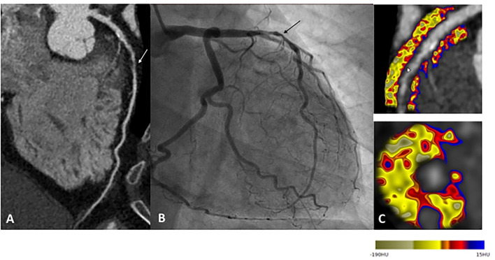
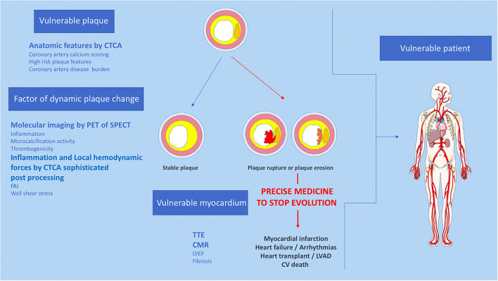
Atherosclerotic plaque rupture or erosion remain the primary mechanism responsible for myocardial infarction and the major challenge of cardiovascular researchers is to develop non-invasive methods of accurate risk prediction to identify vulnerable plaques before the event occurs. Multimodal imaging, by CT-TEP or CT-SPECT, provides both morphological and activity information about the plaque and cumulates the advantages of anatomic and molecular imaging to identify vulnerability features among coronary plaques. However, the rate of acute coronary syndromes remains low and the mechanisms leading to adverse events are clearly more complex than initially assumed. Indeed, recent studies suggest that the detection of a state of vulnerability in a patient is more important than the detection of individual sites of vulnerability as a target of focal treatment. Despite this evolution of concepts, multimodal imaging offers a strong potential to assess patient's vulnerability. Here we review the current state of multimodal imaging to identify vulnerable patients, and then focus on emerging imaging techniques and precision medicine.

摘要

动脉粥样硬化斑块破裂或糜烂仍然是导致心肌梗死的主要机制,心血管研究人员面临的主要挑战是开发非侵入性的准确风险预测方法,以便在事件发生前识别易损斑块。通过CT-TEP或CT-SPECT进行的多模态成像可提供有关斑块的形态学和活性信息,并累积了解剖学和分子成像的优势,以识别冠状动脉斑块中的易损特征。然而,急性冠状动脉综合征的发生率仍然较低,导致不良事件的机制显然比最初设想的更为复杂。事实上,最近的研究表明,检测患者的易损状态比检测作为局部治疗靶点的个体易损部位更为重要。尽管概念有所演变,但多模态成像在评估患者易损性方面具有强大的潜力。在此,我们综述了用于识别易损患者的多模态成像的现状,然后重点介绍新兴的成像技术和精准医学。

https://cdn.ncbi.nlm.nih.gov/pmc/blobs/c46b/8907666/539398c03684/fcvm-09-836473-g0002.jpg
https://cdn.ncbi.nlm.nih.gov/pmc/blobs/c46b/8907666/52cdaf303eac/fcvm-09-836473-g0001.jpg
https://cdn.ncbi.nlm.nih.gov/pmc/blobs/c46b/8907666/539398c03684/fcvm-09-836473-g0002.jpg
https://cdn.ncbi.nlm.nih.gov/pmc/blobs/c46b/8907666/52cdaf303eac/fcvm-09-836473-g0001.jpg
https://cdn.ncbi.nlm.nih.gov/pmc/blobs/c46b/8907666/539398c03684/fcvm-09-836473-g0002.jpg

相似文献

1
Non-invasive Multimodality Imaging of Coronary Vulnerable Patient.冠状动脉易损患者的非侵入性多模态成像
Front Cardiovasc Med. 2022 Feb 24;9:836473. doi: 10.3389/fcvm.2022.836473. eCollection 2022.
2
Vulnerable plaques and patients: state-of-the-art.易损斑块与患者:现状分析。
Eur Heart J. 2020 Aug 14;41(31):2997-3004. doi: 10.1093/eurheartj/ehaa227.
3
Molecular imaging of plaques in coronary arteries with PET and SPECT.正电子发射断层扫描和单光子发射计算机断层扫描在冠状动脉斑块中的分子成像。
J Geriatr Cardiol. 2014 Sep;11(3):259-73. doi: 10.11909/j.issn.1671-5411.2014.03.005.
4
CT Imaging of the Vulnerable Plaque.易损斑块的CT成像
Curr Treat Options Cardiovasc Med. 2017 Nov 6;19(12):92. doi: 10.1007/s11936-017-0592-9.
5
Current Toolset in Predicting Acute Coronary Thrombotic Events: The "Vulnerable Plaque" in a "Vulnerable Patient" Concept.预测急性冠状动脉血栓形成事件的当前工具集:“易损患者”概念中的“易损斑块”
Life (Basel). 2023 Mar 4;13(3):696. doi: 10.3390/life13030696.
6
From vulnerable plaque to vulnerable patient: a call for new definitions and risk assessment strategies: Part II.从易损斑块到易损患者:对新定义和风险评估策略的呼吁:第二部分。
Circulation. 2003 Oct 14;108(15):1772-8. doi: 10.1161/01.CIR.0000087481.55887.C9.
7
Current Advances in the Diagnostic Imaging of Atherosclerosis: Insights into the Pathophysiology of Vulnerable Plaque.动脉粥样硬化的诊断影像学新进展:易损斑块的病理生理学见解。
Int J Mol Sci. 2020 Apr 23;21(8):2992. doi: 10.3390/ijms21082992.
8
From vulnerable plaque to vulnerable patient: a call for new definitions and risk assessment strategies: Part I.从易损斑块到易损患者:呼吁新定义和风险评估策略:第一部分。
Circulation. 2003 Oct 7;108(14):1664-72. doi: 10.1161/01.CIR.0000087480.94275.97.
9
Do plaques rapidly progress prior to myocardial infarction? The interplay between plaque vulnerability and progression.斑块在心肌梗死前是否迅速进展?斑块脆弱性与进展的相互作用。
Circ Res. 2015 Jun 19;117(1):99-104. doi: 10.1161/CIRCRESAHA.117.305637.
10
Early Detection and Treatment of the Vulnerable Coronary Plaque: Can We Prevent Acute Coronary Syndromes?易损冠状动脉斑块的早期检测与治疗:我们能否预防急性冠状动脉综合征?
Circ Cardiovasc Imaging. 2017 May;10(5). doi: 10.1161/CIRCIMAGING.116.005973.

引用本文的文献

1
SPECT-MPI for Coronary Artery Disease: A Deep Learning Approach.用于冠状动脉疾病的单光子发射计算机断层扫描心肌灌注成像:一种深度学习方法。
Acta Med Philipp. 2024 May 15;58(8):67-75. doi: 10.47895/amp.vi0.7582. eCollection 2024.
2
Vulnerable Atherosclerotic Plaque: Is There a Molecular Signature?易损动脉粥样硬化斑块:是否存在分子特征?
Int J Mol Sci. 2022 Nov 7;23(21):13638. doi: 10.3390/ijms232113638.
3
Detecting vulnerable carotid plaque and its component characteristics: Progress in related imaging techniques.检测易损性颈动脉斑块及其成分特征:相关成像技术的进展

本文引用的文献

1
Advances in PET/CT Technology: An Update.PET/CT技术进展:最新情况
Semin Nucl Med. 2022 May;52(3):286-301. doi: 10.1053/j.semnuclmed.2021.10.005. Epub 2021 Nov 22.
2
Artificial Intelligence Based Multimodality Imaging: A New Frontier in Coronary Artery Disease Management.基于人工智能的多模态成像:冠状动脉疾病管理的新前沿。
Front Cardiovasc Med. 2021 Sep 22;8:736223. doi: 10.3389/fcvm.2021.736223. eCollection 2021.
3
Molecular Imaging and Non-molecular Imaging of Atherosclerotic Plaque Thrombosis.动脉粥样硬化斑块血栓形成的分子成像与非分子成像
Front Neurol. 2022 Sep 14;13:982147. doi: 10.3389/fneur.2022.982147. eCollection 2022.
Front Cardiovasc Med. 2021 Jul 5;8:692915. doi: 10.3389/fcvm.2021.692915. eCollection 2021.
4
Spectral photon-counting CT in cardiovascular imaging.心血管成像中的光谱光子计数 CT。
J Cardiovasc Comput Tomogr. 2021 May-Jun;15(3):218-225. doi: 10.1016/j.jcct.2020.12.005. Epub 2020 Dec 21.
5
Colchicine in Patients with Chronic Coronary Disease.秋水仙碱治疗慢性冠心病
N Engl J Med. 2020 Nov 5;383(19):1838-1847. doi: 10.1056/NEJMoa2021372. Epub 2020 Aug 31.
6
Perivascular Fat Attenuation Index Stratifies Cardiac Risk Associated With High-Risk Plaques in the CRISP-CT Study.在CRISP-CT研究中,血管周围脂肪衰减指数对与高危斑块相关的心脏风险进行分层。
J Am Coll Cardiol. 2020 Aug 11;76(6):755-757. doi: 10.1016/j.jacc.2020.05.078.
7
Low-Attenuation Noncalcified Plaque on Coronary Computed Tomography Angiography Predicts Myocardial Infarction: Results From the Multicenter SCOT-HEART Trial (Scottish Computed Tomography of the HEART).冠状动脉计算机断层扫描血管造影中的低衰减非钙化斑块可预测心肌梗死:来自多中心 SCOT-HEART 试验(苏格兰心脏计算机断层扫描)的结果。
Circulation. 2020 May 5;141(18):1452-1462. doi: 10.1161/CIRCULATIONAHA.119.044720. Epub 2020 Mar 16.
8
Will artificial intelligence change the job of the cardiac imaging specialist?人工智能会改变心脏影像专家的工作吗?
Arch Cardiovasc Dis. 2020 Jan;113(1):1-4. doi: 10.1016/j.acvd.2019.11.002. Epub 2019 Dec 30.
9
Efficacy and Safety of Low-Dose Colchicine after Myocardial Infarction.心梗后小剂量秋水仙碱的疗效和安全性。
N Engl J Med. 2019 Dec 26;381(26):2497-2505. doi: 10.1056/NEJMoa1912388. Epub 2019 Nov 16.
10
Expert recommendations on the assessment of wall shear stress in human coronary arteries: existing methodologies, technical considerations, and clinical applications.关于人体冠状动脉壁面剪应力评估的专家建议:现有方法、技术考量及临床应用
Eur Heart J. 2019 Nov 1;40(41):3421-3433. doi: 10.1093/eurheartj/ehz551.