Govil Shrusti Ajay, Asthana Geeta, Kanodia Shikha, Parmar Abhishek
Department of Conservative Dentistry and Endodontics, Government Dental College and Hospital, Ahmedabad, Gujarat, India.
J Conserv Dent. 2021 Jul-Aug;24(4):404-407. doi: 10.4103/JCD.JCD_310_21. Epub 2022 Jan 13.
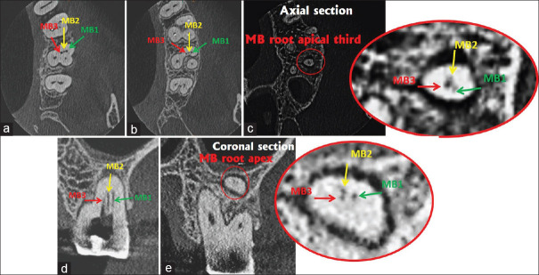
Anatomic variations in mesiobuccal root (MBR) of maxillary molars are common. This variation is found to be more common in maxillary first molars as compared to second molars. However, finding three independent mesiobuccal (MB) canals in the MBR of maxillary molars is clinically a rare entity. With the use of magnification, illumination, and cone-beam computed tomography, combined with the skill of the operator, there is an increased possibility of detecting such additional canals. The present case report describes the successful clinical management of a second molar in a 58-year-old female patient having three MBR canals (MB1, MB2, and MB3) with a Vertucci's Type VIII canal configuration and an almost obliterated pulp chamber. The canals were prepared using hand and rotary instruments, followed by obturation. Very few such cases have been documented clinically in the literature.
上颌磨牙近中颊根(MBR)的解剖变异很常见。与第二磨牙相比,这种变异在上颌第一磨牙中更为常见。然而,在上颌磨牙的MBR中发现三条独立的近中颊(MB)根管在临床上是一种罕见的情况。随着放大、照明和锥形束计算机断层扫描技术的应用,再结合操作者的技能,检测到这种额外根管的可能性增加。本病例报告描述了一名58岁女性患者的第二磨牙的成功临床处理情况,该磨牙有三条MBR根管(MB1、MB2和MB3),呈韦尔图奇VIII型根管形态,且髓室几乎闭塞。使用手动和旋转器械对根管进行预备,随后进行充填。文献中临床记录的此类病例极少。