Thompson Dakota T, Wilkinson Neal, Hrabe Jennifer E, Arshava Evgeny V
Department of Surgery, University of Iowa Carver College of Medicine, 375 Newton Rd, Iowa City, IA 52242, USA.
Department of Surgical Oncology, Kalispell Regional Medical Center, 310 Sunnyview Lane, Kalispell, MT, USA.
Int J Surg Case Rep. 2022 Apr;93:106932. doi: 10.1016/j.ijscr.2022.106932. Epub 2022 Mar 9.
Epidermal inclusion cysts are a common benign finding, and they are predominantly asymptomatic. They can rarely form in the pelvis or abdomen, however, and may cause symptoms secondary to mass effect. This case highlights management of an anterectal epidermal inclusion cyst connected to the perineal cyst, mimicking a dumbbell-shaped lesion, found in a male.
This is a unique case of a 21-year-old Caucasian male with a palpable perineal mass, lower extremity hypoesthesia, and constipation who was found to have a complex-shaped cyst on computed tomography and magnetic resonance imaging. This was ultimately managed with a two-stage perineal and transabdominal resection.
This case highlights that perineal epidermal inclusion cysts may have pelvic extension, especially in patients with additional new-onset neurologic, gastrointestinal, or urologic symptoms. These symptoms should completely resolve after resection. Additionally, resection is recommended to prevent complications including malignant degeneration and fistulization.
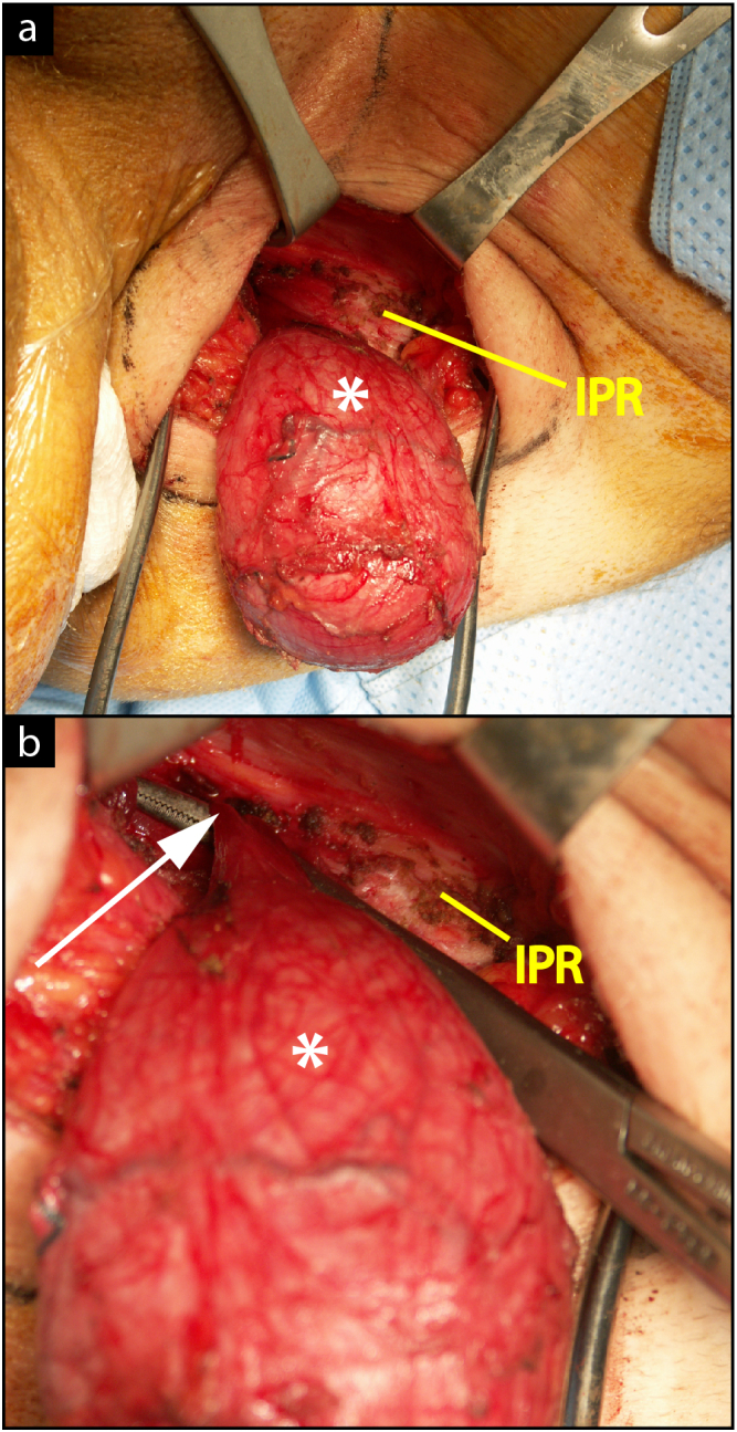
This is the first reported case of an anterectal, epidermal inclusion cyst connected to a perineal cyst found in a male. Perineal and pelvic cysts may be synchronous and may be connected through the pudendal canal. These masses can be safely removed via a combined perineal and transabdominal resection. The connecting portion of lesions that have both pelvic and perineal components should be meticulously identified and dissected because even a thin, patent segment - if left unresected - may result in lesion recurrence.
表皮样囊肿是一种常见的良性病变,多数情况下无症状。然而,它们极少在盆腔或腹部形成,可能因占位效应而引发症状。本病例着重介绍了一名男性患者前直肠表皮样囊肿合并会阴囊肿的处理情况,该囊肿形似哑铃状。
这是一例独特的病例,一名21岁的白种男性,可触及会阴肿物,下肢感觉减退,伴有便秘,经计算机断层扫描和磁共振成像检查发现为复杂形状的囊肿。最终通过两阶段会阴及经腹切除术进行治疗。
该病例表明会阴表皮样囊肿可能向盆腔扩展,尤其是在伴有新发神经、胃肠或泌尿系统症状的患者中。切除术后这些症状应完全缓解。此外,建议进行切除以预防包括恶性变和瘘管形成在内的并发症。
这是首例报道的男性前直肠表皮样囊肿合并会阴囊肿的病例。会阴囊肿和盆腔囊肿可能同时存在,并可能通过阴部管相连。这些肿物可通过会阴联合经腹切除术安全切除。对于同时具有盆腔和会阴部分的病变,其连接部分应仔细识别并解剖,因为即使是细小、通畅的部分——若未切除——也可能导致病变复发。