• 文献检索
  • 文档翻译
  • 深度研究
  • 学术资讯
  • Suppr Zotero 插件Zotero 插件
  • 邀请有礼
  • 套餐&价格
  • 历史记录
应用&插件
Suppr Zotero 插件Zotero 插件浏览器插件Mac 客户端Windows 客户端微信小程序
定价
高级版会员购买积分包购买API积分包
服务
文献检索文档翻译深度研究API 文档MCP 服务
关于我们
关于 Suppr公司介绍联系我们用户协议隐私条款
关注我们

Suppr 超能文献

核心技术专利:CN118964589B侵权必究
粤ICP备2023148730 号-1Suppr @ 2026

文献检索

告别复杂PubMed语法,用中文像聊天一样搜索,搜遍4000万医学文献。AI智能推荐,让科研检索更轻松。

立即免费搜索

文件翻译

保留排版,准确专业,支持PDF/Word/PPT等文件格式,支持 12+语言互译。

免费翻译文档

深度研究

AI帮你快速写综述,25分钟生成高质量综述,智能提取关键信息,辅助科研写作。

立即免费体验

多中心评估在实性不确定小肾肿块中多参数 MRI 透明细胞可能性评分

Multicenter Evaluation of Multiparametric MRI Clear Cell Likelihood Scores in Solid Indeterminate Small Renal Masses.

机构信息

From the Department of Medical Imaging, The Ottawa Hospital, University of Ottawa, Ottawa, Canada (N.S., R.M.H., C.W.); Departments of Radiology (M.S.D., D.B., N.E.C.) and Urology (M.S.D.), University of Michigan Medical Center, Ann Arbor, Mich; Department of Radiology, Brigham and Women's Hospital, Harvard Medical School, Boston, Mass (S.G.S., I.G., A.B.S.); Department of Radiology, NYU Langone Medical Center, New York, NY (B.B., A.M.D., N.H.); Department of Radiology, University of Nebraska Medical Center, Omaha, Neb (Z.B., R.T.D., E.E., E.G.); Department of Epidemiology, Ottawa Hospital Research Institute, Ottawa, Canada (T.R.); and Departments of Radiology and Urology, Advanced Imaging Research Center, University of Texas Southwestern Medical Center, 2201 Inwood Rd, 2nd Floor, Suite 202, Dallas, TX 75390-9085 (I.P.).

出版信息

Radiology. 2022 Jun;303(3):590-599. doi: 10.1148/radiol.211680. Epub 2022 Mar 15.

DOI:10.1148/radiol.211680
PMID:35289659
原文链接:https://pmc.ncbi.nlm.nih.gov/articles/PMC9794383/
Abstract

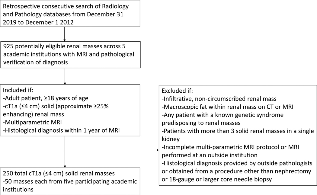
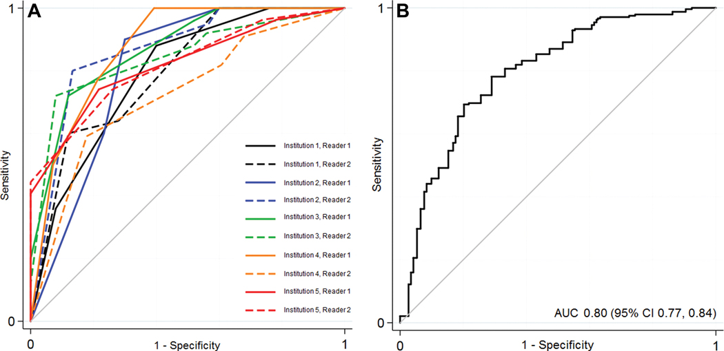
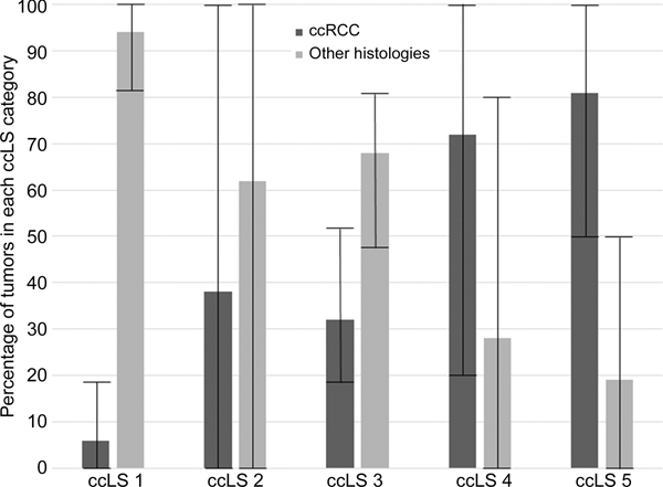
Background Solid small renal masses (SRMs) (≤4 cm) represent benign and malignant tumors. Among SRMs, clear cell renal cell carcinoma (ccRCC) is frequently aggressive. When compared with invasive percutaneous biopsies, the objective of the proposed clear cell likelihood score (ccLS) is to classify ccRCC noninvasively by using multiparametric MRI, but it lacks external validation. Purpose To evaluate the performance of and interobserver agreement for ccLS to diagnose ccRCC among solid SRMs. Materials and Methods This retrospective multicenter cross-sectional study included patients with consecutive solid (≥25% approximate volume enhancement) SRMs undergoing multiparametric MRI between December 2012 and December 2019 at five academic medical centers with histologic confirmation of diagnosis. Masses with macroscopic fat were excluded. After a 1.5-hour training session, two abdominal radiologists per center independently rendered a ccLS for 50 masses. The diagnostic performance for ccRCC was calculated using random-effects logistic regression modeling. The distribution of ccRCC by ccLS was tabulated. Interobserver agreement for ccLS was evaluated with the Fleiss κ statistic. Results A total of 241 patients (mean age, 60 years ± 13 [SD]; 174 men) with 250 solid SRMs were evaluated. The mean size was 25 mm ± 8 (range, 10-39 mm). Of the 250 SRMs, 119 (48%) were ccRCC. The sensitivity, specificity, and positive predictive value for the diagnosis of ccRCC when ccLS was 4 or higher were 75% (95% CI: 68, 81), 78% (72, 84), and 76% (69, 81), respectively. The negative predictive value of a ccLS of 2 or lower was 88% (95% CI: 81, 93). The percentages of ccRCC according to the ccLS were 6% (range, 0%-18%), 38% (range, 0%-100%), 32% (range, 60%-83%), 72% (range, 40%-88%), and 81% (range, 73%-100%) for ccLSs of 1-5, respectively. The mean interobserver agreement was moderate (κ = 0.58; 95% CI: 0.42, 0.75). Conclusion The clear cell likelihood score applied to multiparametric MRI had moderate interobserver agreement and differentiated clear cell renal cell carcinoma from other solid renal masses, with a negative predictive value of 88%. © RSNA, 2022 See also the editorial by Mileto and Potretzke in this issue.

摘要

背景 实体小肾脏肿块(SRMs)(≤4 cm)代表良性和恶性肿瘤。在 SRMs 中,透明细胞肾细胞癌(ccRCC)通常具有侵袭性。与侵入性经皮活检相比,提出的透明细胞可能性评分(ccLS)的目的是通过使用多参数 MRI 对 ccRCC 进行非侵入性分类,但缺乏外部验证。目的 评估 ccLS 用于诊断实体 SRMs 中 ccRCC 的性能和观察者间一致性。材料与方法 本回顾性多中心横断面研究纳入了 2012 年 12 月至 2019 年 12 月在五家学术医疗中心接受多参数 MRI 检查的连续实体(≥25%近似体积增强)SRMs 患者,且经组织学证实诊断。排除有肉眼脂肪的肿块。在 1.5 小时的培训课程后,每个中心的两名腹部放射科医生分别对 50 个肿块进行 ccLS 评分。使用随机效应逻辑回归模型计算 ccRCC 的诊断性能。通过 ccLS 对 ccRCC 进行制表。使用 Fleiss κ 统计量评估 ccLS 的观察者间一致性。结果 共评估了 241 名患者(平均年龄 60 岁±13[SD];174 名男性)的 250 个实体 SRMs。平均大小为 25 mm±8(范围,10-39 mm)。在 250 个 SRMs 中,119 个(48%)为 ccRCC。当 ccLS 为 4 或更高时,ccRCC 诊断的灵敏度、特异性和阳性预测值分别为 75%(95%CI:68,81)、78%(72,84)和 76%(69,81)。ccLS 为 2 或更低时的阴性预测值为 88%(95%CI:81,93)。根据 ccLS,ccRCC 的百分比分别为 6%(范围,0%-18%)、38%(范围,0%-100%)、32%(范围,60%-83%)、72%(范围,40%-88%)和 81%(范围,73%-100%)ccLS 为 1-5。观察者间平均一致性为中度(κ=0.58;95%CI:0.42,0.75)。结论 应用于多参数 MRI 的透明细胞可能性评分具有中度的观察者间一致性,并能区分透明细胞肾细胞癌和其他实体肾肿块,阴性预测值为 88%。©RSNA,2022 另请参见本期 Mileto 和 Potretzke 的社论。

https://cdn.ncbi.nlm.nih.gov/pmc/blobs/e2c3/9794383/23a7fb23db60/nihms-1835684-f0006.jpg
https://cdn.ncbi.nlm.nih.gov/pmc/blobs/e2c3/9794383/57c742c97afb/nihms-1835684-f0001.jpg
https://cdn.ncbi.nlm.nih.gov/pmc/blobs/e2c3/9794383/9bda5d44e305/nihms-1835684-f0002.jpg
https://cdn.ncbi.nlm.nih.gov/pmc/blobs/e2c3/9794383/6023c4763ac3/nihms-1835684-f0003.jpg
https://cdn.ncbi.nlm.nih.gov/pmc/blobs/e2c3/9794383/da38a4335f19/nihms-1835684-f0004.jpg
https://cdn.ncbi.nlm.nih.gov/pmc/blobs/e2c3/9794383/715a9f81588c/nihms-1835684-f0005.jpg
https://cdn.ncbi.nlm.nih.gov/pmc/blobs/e2c3/9794383/23a7fb23db60/nihms-1835684-f0006.jpg

相似文献

1
Multicenter Evaluation of Multiparametric MRI Clear Cell Likelihood Scores in Solid Indeterminate Small Renal Masses.多中心评估在实性不确定小肾肿块中多参数 MRI 透明细胞可能性评分
Radiology. 2022 Jun;303(3):590-599. doi: 10.1148/radiol.211680. Epub 2022 Mar 15.
2
Performance of clear cell likelihood scores in characterizing solid renal masses at multiparametric MRI: an external validation study.透明细胞似然评分在多参数磁共振成像中对实性肾肿块特征描述的性能:一项外部验证研究。
Abdom Radiol (NY). 2023 Mar;48(3):1033-1043. doi: 10.1007/s00261-023-03799-z. Epub 2023 Jan 13.
3
Diagnostic Performance and Interreader Agreement of the MRI Clear Cell Likelihood Score for Characterization of cT1a and cT1b Solid Renal Masses: An External Validation Study.MRI 透明细胞可能性评分对 cT1a 和 cT1b 实性肾脏肿块的特征分析的诊断性能和读者间一致性:一项外部验证研究。
AJR Am J Roentgenol. 2022 Nov;219(5):793-803. doi: 10.2214/AJR.22.27378. Epub 2022 Jun 1.
4
Association of Clear Cell Likelihood Score on MRI and Growth Kinetics of Small Solid Renal Masses on Active Surveillance.磁共振成像上透明细胞可能性评分与主动监测中小肾脏实性肿块生长动力学的关系。
AJR Am J Roentgenol. 2022 Jan;218(1):101-110. doi: 10.2214/AJR.21.25979. Epub 2021 Jul 21.
5
Prospective performance of clear cell likelihood scores (ccLS) in renal masses evaluated with multiparametric magnetic resonance imaging.多参数磁共振成像评估肾肿块中透明细胞可能性评分(ccLS)的前瞻性表现。
Eur Radiol. 2021 Jan;31(1):314-324. doi: 10.1007/s00330-020-07093-0. Epub 2020 Aug 8.
6
Differentiation between renal epithelioid angiomyolipoma and clear cell renal cell carcinoma using clear cell likelihood score.采用透明细胞概率评分鉴别肾上皮样血管平滑肌脂肪瘤与透明细胞肾细胞癌。
Abdom Radiol (NY). 2023 Dec;48(12):3714-3727. doi: 10.1007/s00261-023-04034-5. Epub 2023 Sep 25.
7
Diagnostic performance of prospectively assigned clear cell Likelihood scores (ccLS) in small renal masses at multiparametric magnetic resonance imaging.多参数磁共振成像中前瞻性分配的透明细胞似然评分(ccLS)对小肾肿块的诊断性能
Urol Oncol. 2019 Dec;37(12):941-946. doi: 10.1016/j.urolonc.2019.07.023. Epub 2019 Sep 17.
8
Diagnostic Value of Clear Cell Likelihood Score v1.0 and v2.0 for Common Subtypes of Small Renal Masses: A Multicenter Comparative Study.透明细胞似然评分v1.0和v2.0对小肾肿块常见亚型的诊断价值:一项多中心比较研究
J Magn Reson Imaging. 2025 Jan;61(1):97-110. doi: 10.1002/jmri.29392. Epub 2024 May 13.
9
[Comparison of diagnostic performance of Clear Cell Likelihood Score v1.0 and v2.0 for clear renal cell carcinoma].[透明细胞肾细胞癌的Clear Cell Likelihood Score v1.0和v2.0诊断性能比较]
Nan Fang Yi Ke Da Xue Xue Bao. 2023 May 20;43(5):800-806. doi: 10.12122/j.issn.1673-4254.2023.05.16.
10
Development of a Multiparametric Renal CT Algorithm for Diagnosis of Clear Cell Renal Cell Carcinoma Among Small (≤ 4 cm) Solid Renal Masses.一种用于诊断小(≤ 4cm)实体性肾肿块中透明细胞肾细胞癌的多参数肾 CT 算法的开发。
AJR Am J Roentgenol. 2022 Nov;219(5):814-823. doi: 10.2214/AJR.22.27971. Epub 2022 Jun 29.

引用本文的文献

1
Development and validation of a nomogram combined pre-operative quantitative MR parameters for the prediction of pathological WHO/ISUP grade in clear cell renal cell carcinoma.用于预测透明细胞肾细胞癌病理WHO/ISUP分级的列线图联合术前定量MR参数的开发与验证
World J Urol. 2025 Aug 9;43(1):480. doi: 10.1007/s00345-025-05864-2.
2
Diagnostic performance of the clear cell likelihood score integrated with cystic degeneration or necrosis on MR imaging for identifying clear cell renal cell carcinoma in cT1 solid renal masses.在磁共振成像(MR)上,结合囊性退变或坏死的透明细胞可能性评分对cT1期实性肾肿块中透明细胞肾细胞癌的诊断效能。
Insights Imaging. 2025 Jul 3;16(1):149. doi: 10.1186/s13244-025-02029-y.
3

本文引用的文献

1
How We Do It: Managing the Indeterminate Renal Mass with the MRI Clear Cell Likelihood Score.方法:采用 MRI 透明细胞似然评分管理不确定的肾脏肿块。
Radiology. 2022 Feb;302(2):256-269. doi: 10.1148/radiol.210034. Epub 2021 Dec 14.
2
Active Surveillance of Renal Masses: The Role of Radiology.肾脏肿块的主动监测:放射学的作用。
Radiology. 2022 Jan;302(1):11-24. doi: 10.1148/radiol.2021204227. Epub 2021 Nov 23.
3
New developments in existing WHO entities and evolving molecular concepts: The Genitourinary Pathology Society (GUPS) update on renal neoplasia.
Potential of Metabolic MRI to Address Unmet Clinical Needs in Localised Kidney Cancer.
代谢磁共振成像在满足局限性肾癌未被满足的临床需求方面的潜力。
Cancers (Basel). 2025 May 26;17(11):1773. doi: 10.3390/cancers17111773.
4
Identification of benign from malignant small renal tumors: is there a possible role of T1 mapping?鉴别肾小肿瘤的良恶性:T1 映射成像是否能发挥作用?
Discov Oncol. 2025 May 19;16(1):808. doi: 10.1007/s12672-025-02609-w.
5
In Pursuit of KI-RADS: Toward a Single, Evidence-based Imaging Classification of Renal Masses.追求肾脏影像报告和数据系统(KI-RADS):迈向基于证据的单一肾脏肿块影像分类
Radiology. 2025 Mar;314(3):e240308. doi: 10.1148/radiol.240308.
6
A simple method based on qualitative MRI features for characterizing clear cell renal cell carcinoma in small renal masses: comparison with the clear cell likelihood score.一种基于定性MRI特征的小肾肿块中透明细胞肾细胞癌特征化简单方法:与透明细胞可能性评分的比较
Abdom Radiol (NY). 2025 Feb 19. doi: 10.1007/s00261-025-04844-9.
7
External validation of multiparametric magnetic resonance imaging-based decision rules for characterizing breast lesions and comparison to Kaiser score and breast imaging reporting and data system (BI-RADS) category.基于多参数磁共振成像的乳腺病变特征描述决策规则的外部验证以及与凯泽评分和乳腺影像报告和数据系统(BI-RADS)分类的比较
Quant Imaging Med Surg. 2025 Jan 2;15(1):648-661. doi: 10.21037/qims-23-1783. Epub 2024 Dec 30.
8
'BJUI Clinical Dilemma': the incidental small renal mass in a solitary kidney.《英国泌尿学杂志》临床困境:孤立肾中的偶然发现的小肾肿块
BJU Int. 2025 Mar;135(3):371-379. doi: 10.1111/bju.16627. Epub 2025 Jan 6.
9
Is there enough evidence supporting the clinical adoption of clear cell likelihood score (ccLS)? An updated systematic review and meta-analysis.是否有足够的证据支持临床采用透明细胞可能性评分(ccLS)?一项更新的系统评价和荟萃分析。
Insights Imaging. 2024 Oct 9;15(1):242. doi: 10.1186/s13244-024-01829-y.
10
Diagnostic performance of contrast enhancement to differentiate benign and malignant renal lesions in CT and MRI: a systematic review and meta-analysis of diagnostic test accuracy (DTA) studies.CT和MRI中对比增强鉴别肾脏良恶性病变的诊断性能:诊断试验准确性(DTA)研究的系统评价和荟萃分析
Abdom Radiol (NY). 2025 Jan;50(1):360-378. doi: 10.1007/s00261-024-04514-2. Epub 2024 Aug 13.
现有世卫组织实体的新发展和不断发展的分子概念:泌尿生殖系统病理学学会 (GUPS) 关于肾肿瘤的更新。
Mod Pathol. 2021 Jul;34(7):1392-1424. doi: 10.1038/s41379-021-00779-w. Epub 2021 Mar 4.
4
Prospective performance of clear cell likelihood scores (ccLS) in renal masses evaluated with multiparametric magnetic resonance imaging.多参数磁共振成像评估肾肿块中透明细胞可能性评分(ccLS)的前瞻性表现。
Eur Radiol. 2021 Jan;31(1):314-324. doi: 10.1007/s00330-020-07093-0. Epub 2020 Aug 8.
5
Small Renal Mass Surveillance: Histology-specific Growth Rates in a Biopsy-characterized Cohort.小肾肿物监测:活检特征队列中的组织学特异性生长率
Eur Urol. 2020 Sep;78(3):460-467. doi: 10.1016/j.eururo.2020.06.053. Epub 2020 Jul 14.
6
Diagnostic performance of prospectively assigned clear cell Likelihood scores (ccLS) in small renal masses at multiparametric magnetic resonance imaging.多参数磁共振成像中前瞻性分配的透明细胞似然评分(ccLS)对小肾肿块的诊断性能
Urol Oncol. 2019 Dec;37(12):941-946. doi: 10.1016/j.urolonc.2019.07.023. Epub 2019 Sep 17.
7
Bosniak Classification of Cystic Renal Masses, Version 2019: An Update Proposal and Needs Assessment.Bosniak 分类法在囊性肾脏肿块中的应用(2019 年版):更新建议与需求评估。
Radiology. 2019 Aug;292(2):475-488. doi: 10.1148/radiol.2019182646. Epub 2019 Jun 18.
8
Role of Virtual Biopsy in the Management of Renal Masses.虚拟活检在肾肿物管理中的作用。
AJR Am J Roentgenol. 2019 Jun;212(6):1234-1243. doi: 10.2214/AJR.19.21172. Epub 2019 Apr 17.
9
Interreader Variability of Prostate Imaging Reporting and Data System Version 2 in Detecting and Assessing Prostate Cancer Lesions at Prostate MRI.前列腺影像报告和数据系统第2版在前列腺MRI检测和评估前列腺癌病灶中的阅片者间变异性
AJR Am J Roentgenol. 2019 Jun;212(6):1197-1205. doi: 10.2214/AJR.18.20536. Epub 2019 Mar 27.
10
Update on Indications for Percutaneous Renal Mass Biopsy in the Era of Advanced CT and MRI.先进CT和MRI时代经皮肾肿物活检指征的最新进展
AJR Am J Roentgenol. 2019 Jun;212(6):1187-1196. doi: 10.2214/AJR.19.21093. Epub 2019 Mar 27.