Asan Institute for Life Sciences, Asan Medical Center, University of Ulsan College of Medicine, Seoul, Korea.
Himchan Hospital Bupyeong, Incheon, Korea.
PLoS One. 2022 Mar 24;17(3):e0266030. doi: 10.1371/journal.pone.0266030. eCollection 2022.
Although stem cells might enhance natural enthesis healing in surgical rotator cuff repair, not much attention has been given to the delivery and location of delivering stem cells. The purpose of this study to know where to locate those stem cells during repair.
Animal model of chronic rotator cuff tear was created in 24 rats. Adipose-derived stem cells were engineered as a sheet and transplanted 1) between a torn tendon and humerus (interposition group) or 2) over a repaired tendon-to-bone junction (overlay group) at the time of surgical repair. Tracking of stem cells with overexpression of green fluorescent protein (GFP) were carried out at the time of sacrifice in additional 4 shoulders in each group. Histological and Biomechanical evaluation was performed to compare the differences in tendon-to-bone healing.
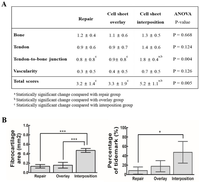
Histology showed increased fibrocartilage, a clear boundary at the mineralized fibrocartilage, abundant collagen type III, and higher total scores, especially in the interposition group. GFP-overexpression was observed at the transplanted site at 2 weeks after repair. Although two groups where stem cell sheets applied showed higher load to failure than the repair-only group, the load to failure was not different between the interposition and overlay group.
In the chronic rotator cuff repair model, stem cell sheets enhanced regeneration of the tendon-to-bone junction. This regeneration was effective when the stem cell sheet was interpositioned at the tendon-to-bone interface.
Basic Science Study; In Vivo Animal Model; Histology and Biomechanics.
尽管干细胞可能会增强手术修复肩袖撕裂后的自然附着愈合,但对于干细胞的输送和定位却没有给予太多关注。本研究旨在了解在修复过程中应将这些干细胞定位在何处。
在 24 只大鼠中建立慢性肩袖撕裂的动物模型。将脂肪来源的干细胞工程化为薄片,并在手术修复时 1)置于撕裂的肌腱和肱骨之间(间置组)或 2)置于修复的肌腱-骨交界处之上(覆盖组)。在每组中的另外 4 个肩膀上进行过表达绿色荧光蛋白(GFP)的干细胞追踪。进行组织学和生物力学评估,以比较肌腱-骨愈合的差异。
组织学显示纤维软骨增加,矿化纤维软骨有明确的边界,富含 III 型胶原,总评分较高,间置组尤其明显。在修复后 2 周,在移植部位观察到 GFP 过表达。尽管应用干细胞片的两组的失效负荷均高于仅修复组,但间置组和覆盖组之间的失效负荷没有差异。
在慢性肩袖修复模型中,干细胞片增强了肌腱-骨交界处的再生。当将干细胞片置于肌腱-骨界面之间时,这种再生是有效的。
基础科学研究;体内动物模型;组织学和生物力学。