Department of Neurology, Medical Faculty Mannheim and Mannheim Center of Translational Neurosciences (MCTN), Heidelberg University, Theodor-Kutzer-Ufer 1-3, 68167, Mannheim, Germany.
Institute for Innate Immunoscience, Medical Faculty Mannheim, Heidelberg University, Mannheim, Germany.
J Neurol. 2022 Aug;269(8):4414-4420. doi: 10.1007/s00415-022-11082-2. Epub 2022 Mar 25.
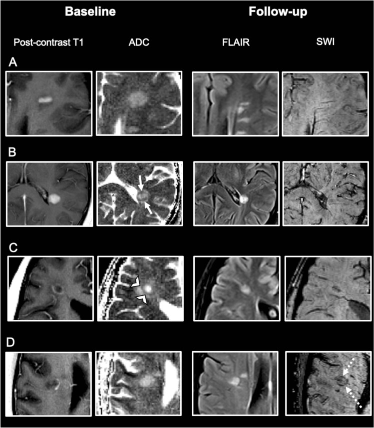
In multiple sclerosis (MS), iron rim lesions (IRLs) are characterized by progressive tissue matrix damage. Therefore, early identification could represent an interesting target for therapeutic intervention to minimize evolving tissue damage. The aim of this study was to identify magnetic resonance imaging (MRI) parameters predicting the conversion from contrast-enhancing to IRLs.
We retrospective identified MS patients scanned on the same 3 T MRI system presenting at least one supratentorial contrast-enhancing lesion (CEL) and a second MRI including susceptibility-weighted images after at least 3 months. On baseline MRI, pattern of contrast-enhancement was categorized as "nodular" or "ring-like", apparent diffusion coefficient (ADC) maps were assessed for the presence of a peripheral hypointense rim. Lesion localization, quantitative volumes (ADC, lesion volume) and the presence of a central vein were assessed.
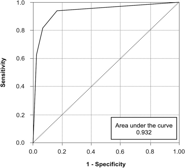
Eighty-nine acute contrast-enhancing lesions in 54 MS patients were included. On follow-up, 16/89 (18%) initially CELs converted into IRLs. CELs that converted into IRLs were larger and demonstrated significantly more often a ring-like contrast-enhancement pattern and a peripheral hypointense rim on ADC maps. Logistic regression model including the covariables pattern of contrast-enhancement and presence of a hypointense rim on ADC maps showed the best predictive performance (area under the curve = 0.932).
The combination of a ring-like contrast-enhancement pattern and a peripheral hypointense rim on ADC maps has the ability to predict the evolution from acute to IRLs. This could be of prognostic value and become a target for early therapeutic intervention to minimize the associated tissue damage.
在多发性硬化症(MS)中,铁环病变(IRL)的特征是进行性组织基质损伤。因此,早期识别可能是治疗干预的一个有趣目标,以最大限度地减少正在进行的组织损伤。本研究的目的是确定预测从增强对比到 IRL 转变的磁共振成像(MRI)参数。
我们回顾性地确定了在同一 3 T MRI 系统上扫描的 MS 患者,这些患者至少有一个幕上增强对比病变(CEL)和第二次 MRI,至少在 3 个月后包括磁化率加权图像。在基线 MRI 上,对比增强的模式分为“结节状”或“环状”,评估表观扩散系数(ADC)图是否存在外周低信号环。评估病变定位、定量体积(ADC、病变体积)和中央静脉的存在。
54 例 MS 患者的 89 个急性增强病变被纳入研究。在随访中,16/89(18%)最初的 CEL 转化为 IRL。转化为 IRL 的 CEL 更大,在 ADC 图上更常显示出环状对比增强模式和外周低信号环。包括对比增强模式和 ADC 图上低信号环存在这两个协变量的逻辑回归模型显示出最佳的预测性能(曲线下面积=0.932)。
环状对比增强模式和 ADC 图上外周低信号环的结合具有预测从急性到 IRL 演变的能力。这可能具有预后价值,并成为早期治疗干预的目标,以最大限度地减少相关的组织损伤。