National Key Laboratory of Medical Immunology & Institute of Immunology , Second Military Medical University , Shanghai , China.
Center for Immunotherapy , Chinese Academy of Medical Sciences , Beijing , China.
Hepatology. 2023 Apr 1;77(4):1106-1121. doi: 10.1002/hep.32487. Epub 2022 Apr 11.
Hepatocarcinogenesis goes through HCC progenitor cells (HcPCs) to fully established HCC, and the mechanisms driving the development of HcPCs are still largely unknown.
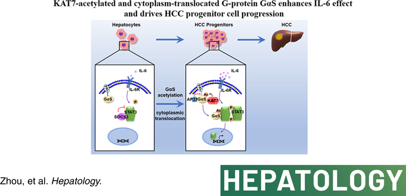
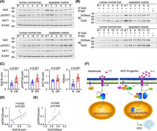
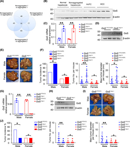
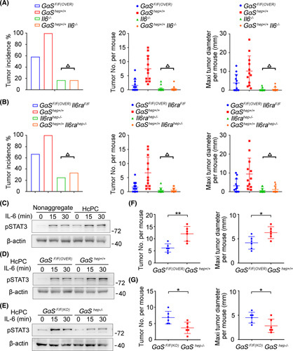
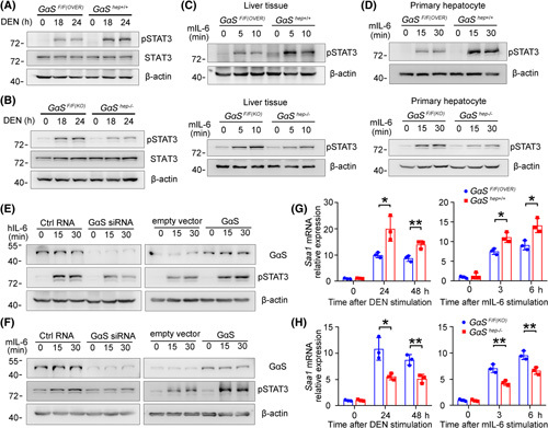
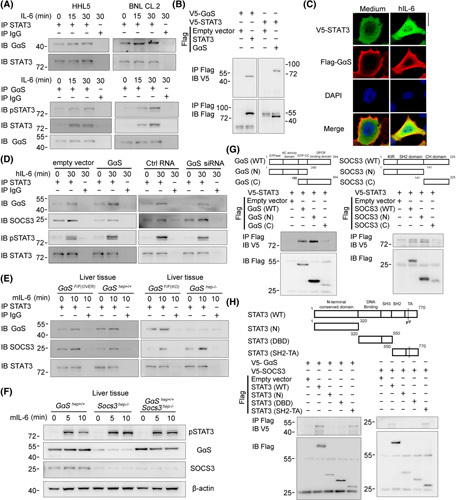
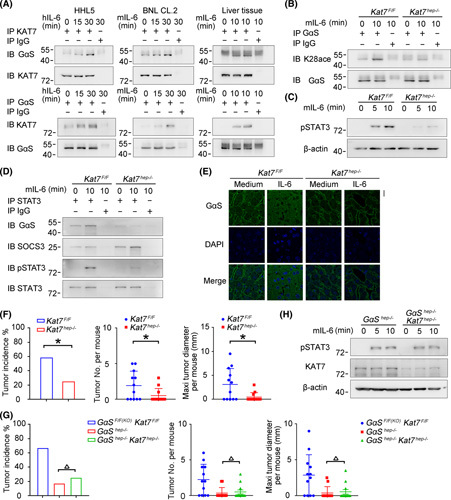
Proteomic analysis in nonaggregated hepatocytes and aggregates containing HcPCs from a diethylnitrosamine-induced HCC mouse model was screened using a quantitative mass spectrometry-based approach to elucidate the dysregulated proteins in HcPCs. The heterotrimeric G stimulating protein α subunit (GαS) protein level was significantly increased in liver cancer progenitor HcPCs, which promotes their response to oncogenic and proinflammatory cytokine IL-6 and drives premalignant HcPCs to fully established HCC. Mechanistically, GαS was located at the membrane inside of hepatocytes and acetylated at K28 by acetyltransferase lysine acetyltransferase 7 (KAT7) under IL-6 in HcPCs, causing the acyl protein thioesterase 1-mediated depalmitoylation of GαS and its cytoplasmic translocation, which were determined by GαS K28A mimicking deacetylation or K28Q mimicking acetylation mutant mice and hepatic Kat7 knockout mouse. Then, cytoplasmic acetylated GαS associated with signal transducer and activator of transcription 3 (STAT3) to impede its interaction with suppressor of cytokine signaling 3, thus promoting in a feedforward manner STAT3 phosphorylation and the response to IL-6 in HcPCs. Clinically, GαS, especially K28-acetylated GαS, was determined to be increased in human hepatic premalignant dysplastic nodules and positively correlated with the enhanced STAT3 phosphorylation, which were in accordance with the data obtained in mouse models.
Malignant progression of HcPCs requires increased K28-acetylated and cytoplasm-translocated GαS, causing enhanced response to IL-6 and driving premalignant HcPCs to fully established HCC, which provides mechanistic insight and a potential target for preventing hepatocarcinogenesis.
肝癌发生经过 HCC 祖细胞(HcPCs)到完全建立的 HCC,驱动 HcPCs 发展的机制在很大程度上仍然未知。
采用基于定量质谱的方法对二乙基亚硝胺诱导的 HCC 小鼠模型中非聚集的肝细胞和含有 HcPCs 的聚集物进行蛋白质组学分析,以阐明 HcPCs 中失调的蛋白质。在肝癌祖细胞 HcPCs 中,异三聚体 G 刺激蛋白α亚基(GαS)蛋白水平显著增加,促进其对致癌和促炎细胞因子 IL-6 的反应,并驱动前恶性 HcPCs 发展为完全建立的 HCC。在机制上,GαS 位于肝细胞内的膜上,在 HcPCs 中的 IL-6 作用下被乙酰转移酶赖氨酸乙酰转移酶 7(KAT7)乙酰化在 K28 上,导致酰基蛋白硫酯酶 1 介导的 GαS 去棕榈酰化及其细胞质易位,这是由 GαS K28A 模拟去乙酰化或 K28Q 模拟乙酰化突变小鼠和肝 Kat7 敲除小鼠确定的。然后,细胞质乙酰化的 GαS 与信号转导和转录激活因子 3(STAT3)结合,阻止其与细胞因子信号转导抑制因子 3 相互作用,从而以正反馈方式促进 STAT3 磷酸化和 HcPCs 对 IL-6 的反应。临床上,GαS,特别是 K28 乙酰化的 GαS,在人类肝前恶性发育不良结节中被确定增加,并且与增强的 STAT3 磷酸化呈正相关,这与在小鼠模型中获得的数据一致。
HcPCs 的恶性进展需要增加的 K28 乙酰化和细胞质易位的 GαS,导致对 IL-6 的增强反应并驱动前恶性 HcPCs 发展为完全建立的 HCC,这为预防肝癌发生提供了机制见解和潜在的靶标。