Sorbonne Université, INSERM, UMR_S 1166, Faculté de Médecine Pitié-Salpêtrière Paris France.
ICAN Institute Paris France.
J Am Heart Assoc. 2022 Apr 5;11(7):e023021. doi: 10.1161/JAHA.121.023021. Epub 2022 Mar 29.
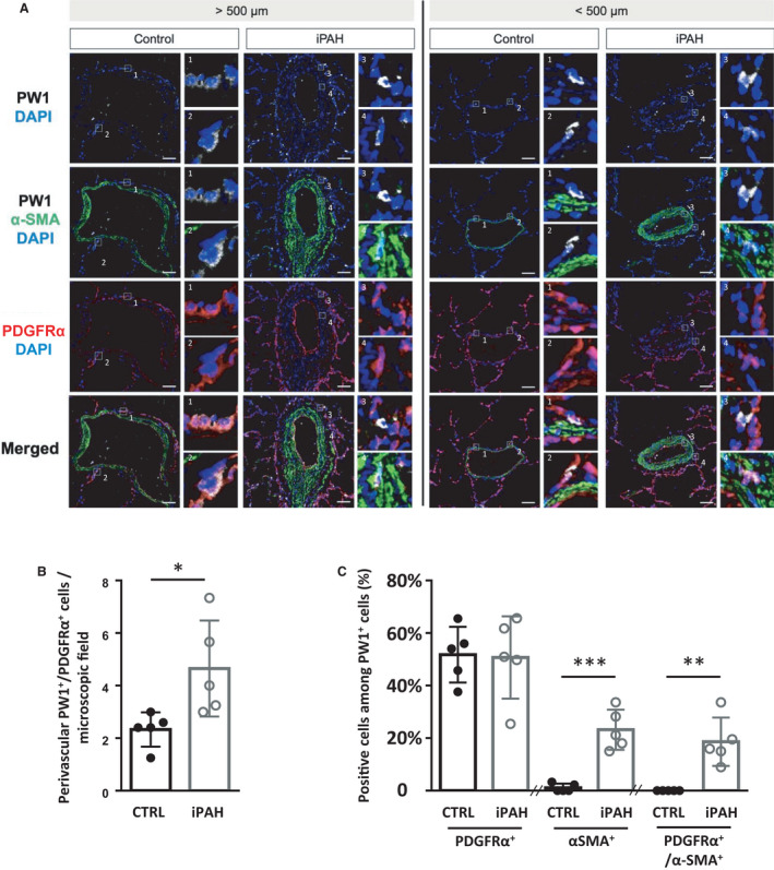
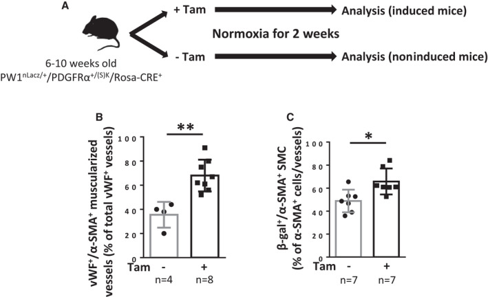
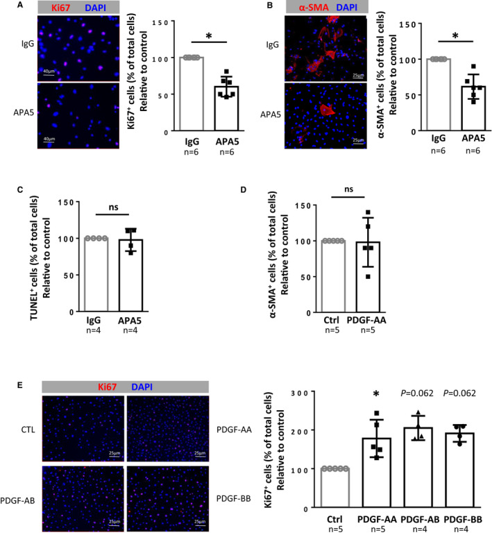
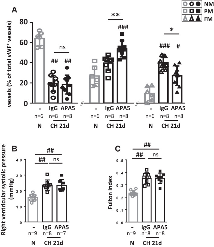
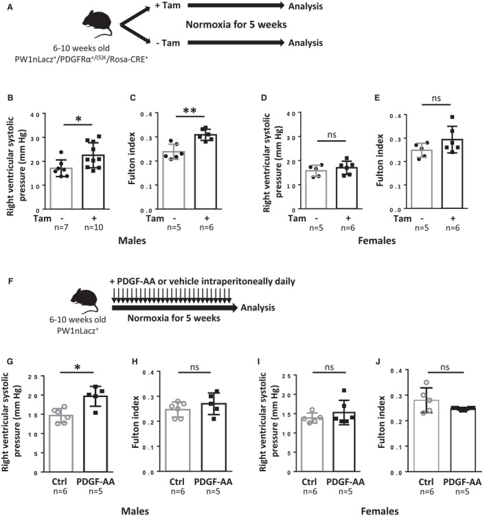
Background Platelet-derived growth factor is a major regulator of the vascular remodeling associated with pulmonary arterial hypertension. We previously showed that protein widely 1 (PW1) vascular progenitor cells participate in early vessel neomuscularization during experimental pulmonary hypertension (PH) and we addressed the role of the platelet-derived growth factor receptor type α (PDGFRα) pathway in progenitor cell-dependent vascular remodeling and in PH development. Methods and Results Remodeled pulmonary arteries from patients with idiopathic pulmonary arterial hypertension showed an increased number of perivascular and vascular PW1 cells expressing PDGFRα. PW1 reporter mice were used to follow the fate of pulmonary PW1 progenitor cells in a model of chronic hypoxia-induced PH development. Under chronic hypoxia, PDGFRα inhibition prevented the increase in PW1 progenitor cell proliferation and differentiation into vascular smooth muscle cells and reduced pulmonary vessel neomuscularization, but did not prevent an increased right ventricular systolic pressure or the development of right ventricular hypertrophy. Conversely, constitutive PDGFRα activation led to neomuscularization via PW1 progenitor cell differentiation into new smooth muscle cells and to PH development in male mice without fibrosis. In vitro, PW1 progenitor cell proliferation, but not differentiation, was dependent on PDGFRα activity. Conclusions These results demonstrate a major role of PDGFRα signaling in progenitor cell-dependent lung vessel neomuscularization and vascular remodeling contributing to PH development, including in idiopathic pulmonary arterial hypertension patients. Our findings suggest that PDGFRα blockers may offer a therapeutic add-on strategy to combine with current pulmonary arterial hypertension treatments to reduce vascular remodeling. Furthermore, our study highlights constitutive PDGFRα activation as a novel experimental PH model.
背景 血小板衍生生长因子是与肺动脉高压相关的血管重构的主要调节因子。我们之前曾表明,广泛表达蛋白 1(PW1)的血管祖细胞参与实验性肺动脉高压(PH)早期的血管新生肌化,我们探讨了血小板衍生生长因子受体α(PDGFRα)途径在祖细胞依赖性血管重构和 PH 发展中的作用。
方法和结果 特发性肺动脉高压患者的重构肺动脉表现出血管周和血管 PW1 细胞表达 PDGFRα 的数量增加。使用 PW1 报告小鼠来追踪慢性低氧诱导的 PH 发展模型中肺 PW1 祖细胞的命运。在慢性低氧下,PDGFRα 抑制可防止 PW1 祖细胞增殖和分化为血管平滑肌细胞增加,并减少肺血管新生肌化,但不能防止右心室收缩压升高或右心室肥厚的发展。相反,组成型 PDGFRα 激活通过 PW1 祖细胞分化为新的平滑肌细胞导致新生肌化,并导致雄性小鼠没有纤维化的 PH 发展。在体外,PW1 祖细胞的增殖,但不是分化,依赖于 PDGFRα 活性。
结论 这些结果表明 PDGFRα 信号在祖细胞依赖性肺血管新生肌化和血管重构中起主要作用,有助于 PH 的发展,包括特发性肺动脉高压患者。我们的研究结果表明,PDGFRα 阻滞剂可能提供一种治疗附加策略,与当前的肺动脉高压治疗相结合,以减少血管重构。此外,我们的研究强调了组成型 PDGFRα 激活作为一种新的实验性 PH 模型。