Filippetti Laura, Pace Nathalie, Louis Jean-Sebastien, Mandry Damien, Goehringer François, Rocher Maria-Soledad, Jay Nicolas, Selton-Suty Christine, Hossu Gabriela, Huttin Olivier, Marie Pierre-Yves
Department of Cardiology, CHRU-Nancy, Nancy, France.
Université de Lorraine, INSERM, UMR-1254, Nancy, France.
Front Cardiovasc Med. 2022 Mar 9;9:831580. doi: 10.3389/fcvm.2022.831580. eCollection 2022.
This observational CMR study aims to characterize left-ventricular (LV) damage, which may be specifically attributed to COVID-19 and is distant in time from the acute phase, through serial CMR performed during the first year in patients with no prior cardiac disease.
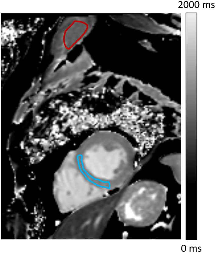
This study included consecutive patients without any prior history of cardiac disease but with a peak troponin-Ic > 50 ng/ml at the time of the first COVID-wave. All had a CMR in the first months after the acute phase, and some had an additional CMR at the end of the first year to monitor LV function, remodeling, and abnormalities evocative of myositis and myocarditis - i.e., increased T1/T2 relaxation times, increased extracellular volume (ECV), and delayed contrast enhancement.
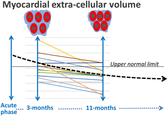
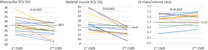
Nineteen consecutively admitted COVID-19 patients (17 men, median age 66 [57-71] years) were included. Eight (42%) had hypertension, six (32%) were obese, and 16 (84%) had suffered an acute respiratory distress syndrome. The 1 CMR, recorded at a median 3.2 [interquartile range: 2.6-3.9] months from the troponin peak, showed (1) LV concentric remodeling in 12 patients (63%), (2) myocardial tissue abnormalities in 11 (58%), including 9 increased myocardial ECVs, and (3) 14 (74%) increased ECVs from shoulder skeletal muscles. The 2 CMR, obtained at 11.1 [11.0-11.7] months from the troponin peak in 13 patients, showed unchanged LV function and remodeling but a return to normal or below the normal range for all ECVs of the myocardium and skeletal muscles.
Many patients with no history of cardiac disease but for whom an increase in blood troponin-Ic ascertained COVID-19 induced myocardial damage exhibited signs of persistent extracellular edema at a median 3-months from the troponin peak, affecting the myocardium and skeletal muscles, which resolved within a one-year time frame. Associations with long-COVID symptoms need to be investigated on a larger scale now.
NCT04753762 on the ClinicalTrials.gov site.
这项观察性心脏磁共振成像(CMR)研究旨在通过对无心脏病史患者在第一年进行的系列CMR检查,来描述可能明确归因于新型冠状病毒肺炎(COVID-19)且与急性期时间间隔较长的左心室(LV)损伤特征。
本研究纳入了连续的无任何心脏病史但在第一波COVID疫情期间肌钙蛋白Ic峰值>50 ng/ml的患者。所有患者在急性期后的头几个月均接受了CMR检查,部分患者在第一年末还进行了额外的CMR检查,以监测左心室功能、重塑以及提示肌炎和心肌炎的异常情况,即T1/T2弛豫时间增加、细胞外容积(ECV)增加和延迟强化。
纳入了19例连续收治的COVID-19患者(17例男性,中位年龄66 [57 - 71]岁)。8例(42%)患有高血压,6例(32%)肥胖,16例(84%)曾发生急性呼吸窘迫综合征。首次CMR检查在距肌钙蛋白峰值中位数3.2 [四分位间距:2.6 - 3.9]个月时进行,结果显示:(1)12例患者(63%)出现左心室向心性重塑;(2)11例患者(58%)存在心肌组织异常,其中9例心肌ECV增加;(3)14例患者(74%)肩部骨骼肌ECV增加。13例患者在距肌钙蛋白峰值11.1 [11.0 - 11.7]个月时进行的第二次CMR检查显示,左心室功能和重塑未改变,但心肌和骨骼肌的所有ECV均恢复至正常或低于正常范围。
许多无心脏病史但血液中肌钙蛋白Ic升高确诊为COVID-19所致心肌损伤的患者,在距肌钙蛋白峰值中位数3个月时表现出持续性细胞外水肿的迹象,累及心肌和骨骼肌,且在1年时间内消退。目前需要更大规模地研究其与长新冠症状的关联。
ClinicalTrials.gov网站上的NCT04753762。