Department of Radiology, University Hospital Muenster, University of Muenster, D-48149 Muenster, Germany.
Department of Radiology, University Hospital Muenster, University of Muenster, D-48149 Muenster, Germany.
Neoplasia. 2022 Jun;28:100792. doi: 10.1016/j.neo.2022.100792. Epub 2022 Mar 31.
As a promotor of tumor invasion and tumor microenvironment (TME) formation, the protein complex S100A8/S100A9 is associated with poor prognosis. Our aim was to further evaluate its origin and regulatory effects, and to establish an imaging biomarker for TME activity.
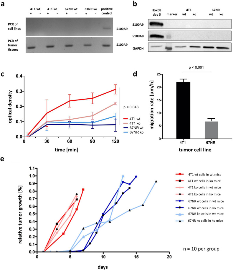
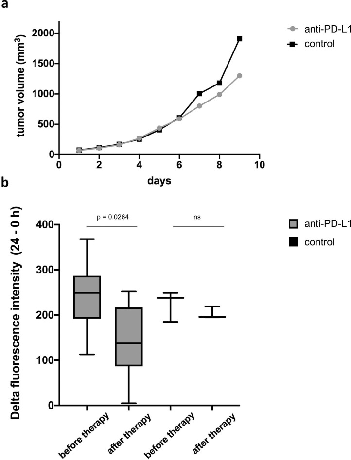
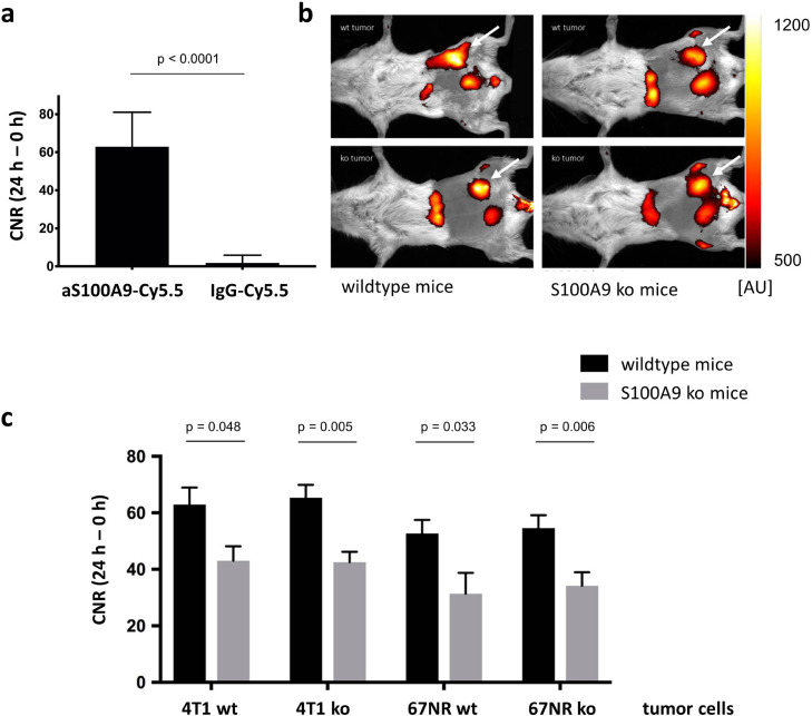
S100A9cells (ko) were created from syngeneic murine breast cancer 4T1 (high malignancy) and 67NR (low malignancy) wildtype (wt) cell lines and implanted into either female BALB/c wildtype or S100A9 mice (n = 10 each). Anti-S100A9-Cy5.5-targeted fluorescence reflectance imaging was performed at 0 h and 24 h after injection. Potential early changes of S100A9-presence under immune checkpoint inhibition (anti-PD-L1, n = 7 vs. rat IgG2b as isotype control, n = 3) were evaluated.
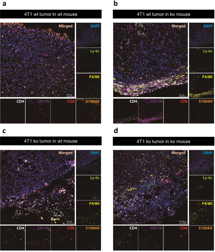
In S100A9mice contrast-to-noise-ratios were significantly reduced for wt and S100A9tumors. No significant differences were detected for 4T1 ko and 67NR ko cells as compared to wildtype cells. Under anti-PD-L1 treatment S100A9 presence significantly decreased compared with the control group.
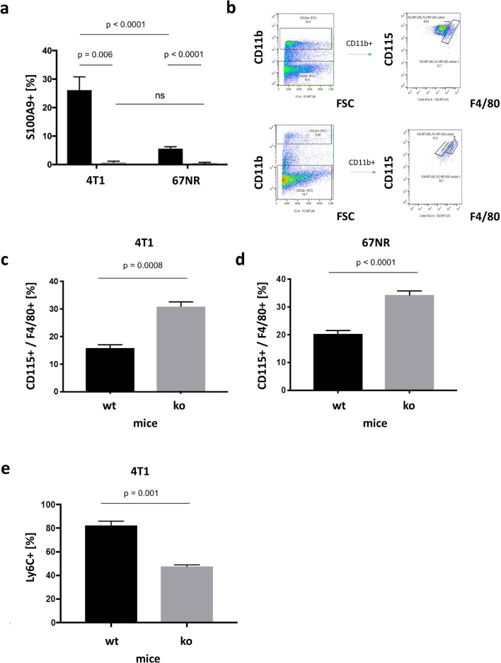
Our results confirm a secretion of S100A8/S100A9 by the TME, while tumor cells do not apparently release the protein. Under immune checkpoint inhibition S100A9-imaging reports an early decrease of TME activity. Therefore, S100A9-specific imaging may serve as an imaging biomarker for TME formation and activity.
作为肿瘤侵袭和肿瘤微环境(TME)形成的促进剂,S100A8/S100A9 蛋白复合物与预后不良相关。我们的目的是进一步评估其来源和调节作用,并建立 TME 活性的成像生物标志物。
从同源小鼠乳腺癌 4T1(高恶性)和 67NR(低恶性)野生型(wt)细胞系中创建 S100A9 细胞(ko),并将其植入雌性 BALB/c 野生型或 S100A9 小鼠(每组 10 只)中。在注射后 0 h 和 24 h 进行抗 S100A9-Cy5.5 靶向荧光反射成像。评估免疫检查点抑制(抗 PD-L1,n=7 与大鼠 IgG2b 作为同型对照,n=3)下 S100A9 存在的潜在早期变化。
在 S100A9 小鼠中,wt 和 S100A9 肿瘤的对比噪声比显着降低。与野生型细胞相比,4T1 ko 和 67NR ko 细胞未检测到显着差异。与对照组相比,在抗 PD-L1 治疗下,S100A9 的存在显着减少。
我们的结果证实了 TME 分泌 S100A8/S100A9,而肿瘤细胞显然不会释放该蛋白。在免疫检查点抑制下,S100A9 成像报告 TME 活性的早期下降。因此,S100A9 特异性成像可作为 TME 形成和活性的成像生物标志物。