Division of Collaboration and Education, International Institute for Zoonosis Control, Hokkaido University, Sapporo, Japan.
Department of Pathology, Albert Einstein College of Medicine, Bronx, NY, United States.
Front Cell Infect Microbiol. 2022 Mar 14;12:848693. doi: 10.3389/fcimb.2022.848693. eCollection 2022.
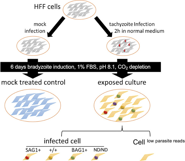
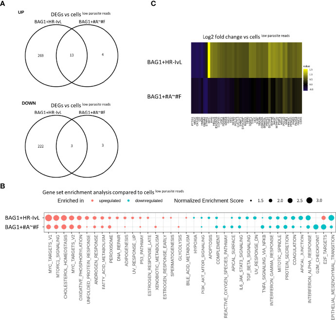
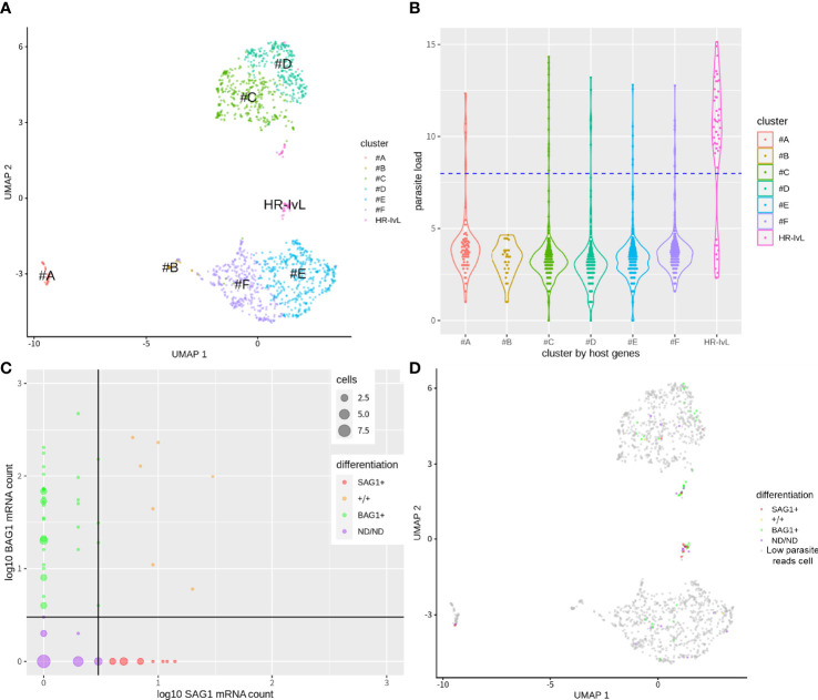
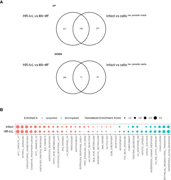
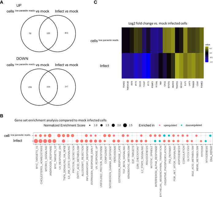
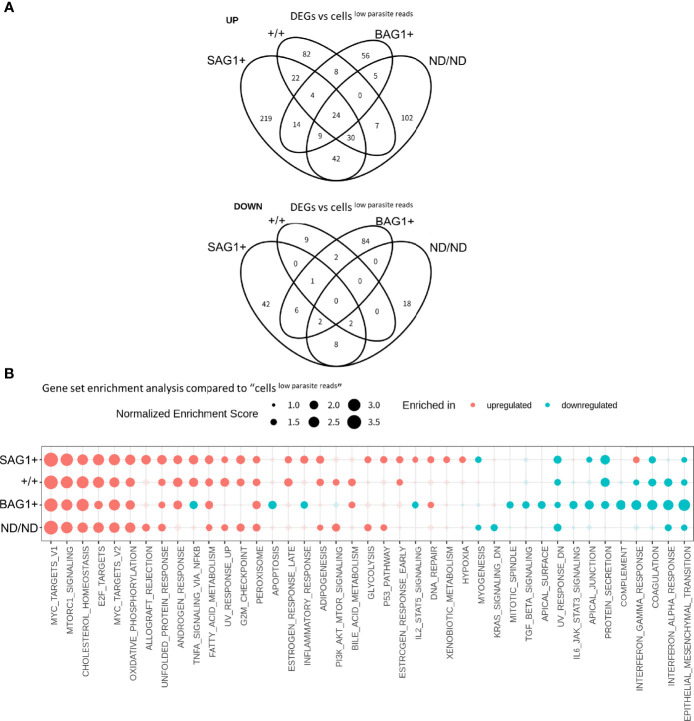
bradyzoites establish chronic infections within their host cells. Recent studies have demonstrated that several parasite effector proteins are translocated to host cells during the bradyzoite stage of chronic infection. To understand the interaction between host cells and bradyzoites at the transcriptomic landscape level, we utilized single-cell RNA-sequencing (scRNA-Seq) to characterize the bradyzoite-induced host cell response. Distinct gene expression profiles were observed in infected host, cells with low parasite mapped reads, and mock (non-exposed) control cells. Gene set enrichment analysis showed that c-Myc and NF-κB signaling and energy metabolic pathways were upregulated by infection. Type I and II interferon response pathways were upregulated in cells with low parasite mapped reads compared to the non-exposed host control cells, and this upregulation effect was reversed in infected cells. Differences were observed in the host cells depending on the differentiation status of the parasites, as determined by BAG1 and SAG1 expression. NF-κB, inflammatory response pathways, and IFN-γ response pathways were downregulated in host cells containing , whereas this downregulation effect was reversed in case of . We also identified two distinct host cell subsets that contained , one of which displayed distinct transcriptomes with upregulated c-Myc expression. Overall, these data clearly demonstrate that host cell transcriptional alteration by bradyzoite infection is different from that of tachyzoite infection, indicating fine-tuning of the host immune response.
缓殖子在宿主细胞内建立慢性感染。最近的研究表明,几种寄生虫效应蛋白在慢性感染的缓殖子阶段被转运到宿主细胞中。为了在转录组景观水平上了解宿主细胞与缓殖子之间的相互作用,我们利用单细胞 RNA 测序 (scRNA-Seq) 来描述缓殖子诱导的宿主细胞反应。在受感染的宿主细胞、寄生虫映射读数低的细胞和模拟(未暴露)对照细胞中观察到不同的基因表达谱。基因集富集分析表明,感染导致 c-Myc 和 NF-κB 信号转导以及能量代谢途径上调。与未暴露的宿主对照细胞相比,寄生虫映射读数低的细胞中 I 型和 II 型干扰素反应途径上调,而感染细胞中的这种上调作用被逆转。根据 BAG1 和 SAG1 表达,宿主细胞因寄生虫的分化状态而存在差异。NF-κB、炎症反应途径和 IFN-γ 反应途径在含有 的宿主细胞中下调,而在 的情况下,这种下调作用被逆转。我们还鉴定了两种含有 的宿主细胞亚群,其中一种显示出 c-Myc 表达上调的独特转录组。总体而言,这些数据清楚地表明,缓殖子感染引起的宿主细胞转录改变与速殖子感染不同,表明宿主免疫反应的精细调控。