Department of Gynecology, Obstetrics and Reproductive Medicine, University Medical School of Saarland, Kirrberger Straße, 66421, Homburg/Saar, Germany.
Institute of Medical Biometry, Epidemiology and Medical Informatics, Saarland University, 66421, Homburg, Germany.
Arch Gynecol Obstet. 2022 Nov;306(5):1689-1695. doi: 10.1007/s00404-022-06529-w. Epub 2022 Apr 4.
PD-L1 receptor expression in breast cancer tissue can be assessed with different anti-human PD-L1 monoclonal antibodies. The performance of three specific monoclonal antibodies in a head-to-head comparison is unknown. In addition, a potential correlation of PD-L1 expression and clinico-pathological parameters has not been investigated.
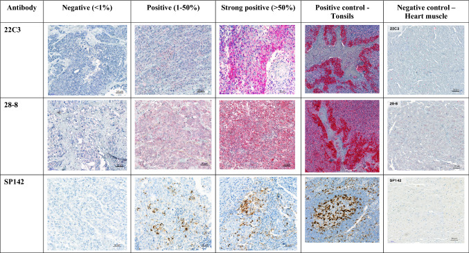
This was a retrospective study on tissue samples of patients with histologically confirmed triple negative breast cancer (TNBC). PD-L1 receptors were immune histochemically stained with three anti-human PD-L1 monoclonal antibodies: 22C3 and 28-8 for staining of tumor cell membranes (TC) and cytoplasm (Cyt), SP142 for immune cell staining (IC). Three different tissue samples of each patient were evaluated separately by two observers in a blinded fashion. The percentage of PD-L1 positive tumor cells in relation to the total number of tumor cells was determined. For antibodies 22C3 and 28-8 PD-L1 staining of 0 to < 1% of tumor cells was rated "negative", 1-50% was rated "positive" and > 50% was rated "strong positive". Cyt staining was defined as "negative" when no signal was observed and as "positive", when any positive signal was observed. For IC staining with SP142 all samples with PD-L1 expression ≥ 1% were rated as "positive". Finally, the relationship between PD-L1 expression and clinico-pathological parameters was analyzed.
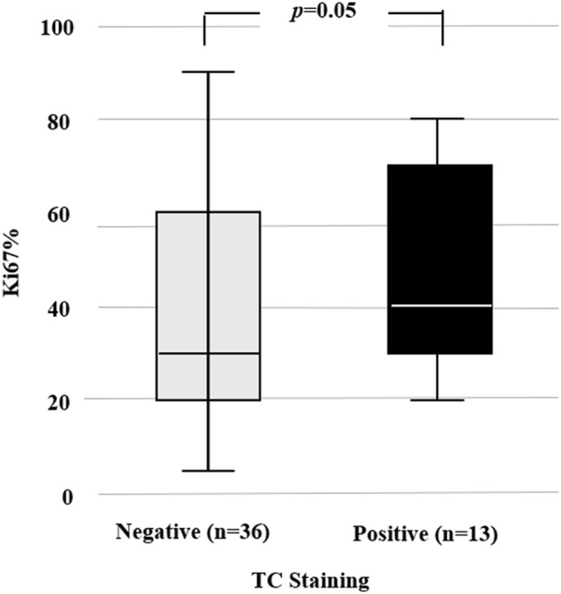
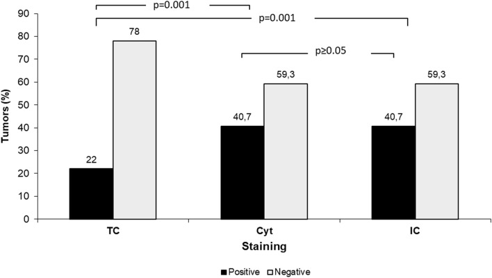
Tissue samples from 59 of 60 enrolled patients could be analyzed. Mean age was 55 years. Both the monoclonal antibodies 22C3 and 28-8 had similar properties, and were positive for both TC in 13 patients (22%) and for Cyt staining in 24 patients (40.7%). IC staining with antibody SP142 was positive in 24 patients (40.7%), who were also positive for Cyt staining. The differences between TC and Cyt staining and TC and IC staining were significant (p = 0.001). Cases with positive TC staining showed higher Ki67 expression compared to those with negative staining, 40 vs 30%, respectively (p = 0.05). None of the other clinico-pathological parameters showed any correlation with PDL1 expression.
Antibodies 22C3 and 28-8 can be used interchangeably for PD-L1 determination in tumor cells of TNBC patients. Results for Cyt staining with 22C3 or 28-8 and IC staining with SP142 were identical. In our study PD-L1 expression correlates with Ki67 expression but not with OS or DFS.
不同的抗人 PD-L1 单克隆抗体可用于评估乳腺癌组织中的 PD-L1 受体表达。三种特定单克隆抗体的性能在头对头比较中尚不清楚。此外,PD-L1 表达与临床病理参数之间的潜在相关性尚未得到研究。
这是一项对组织学证实为三阴性乳腺癌(TNBC)患者的组织样本进行的回顾性研究。使用三种抗人 PD-L1 单克隆抗体:22C3 和 28-8 用于染色肿瘤细胞膜(TC)和细胞质(Cyt),SP142 用于免疫细胞染色(IC),对 PD-L1 受体进行免疫组织化学染色。由两名观察者以盲法方式分别对每位患者的三种不同组织样本进行评估。根据总肿瘤细胞数确定 PD-L1 阳性肿瘤细胞的百分比。对于抗体 22C3 和 28-8,将肿瘤细胞<1%的 PD-L1 染色评为“阴性”,1-50%评为“阳性”,>50%评为“强阳性”。当未观察到信号时,Cyt 染色被定义为“阴性”,当观察到任何阳性信号时,被定义为“阳性”。对于 SP142 的 IC 染色,所有 PD-L1 表达≥1%的样本均评为“阳性”。最后,分析 PD-L1 表达与临床病理参数之间的关系。
纳入的 60 例患者中有 59 例的组织样本可进行分析。平均年龄为 55 岁。单克隆抗体 22C3 和 28-8 的性质相似,13 例(22%)患者 TC 阳性,24 例(40.7%)Cyt 染色阳性。抗体 SP142 的 IC 染色阳性 24 例(40.7%),同时也有 Cyt 染色阳性。TC 与 Cyt 染色以及 TC 与 IC 染色之间的差异具有统计学意义(p=0.001)。TC 染色阳性的病例与 TC 染色阴性的病例相比,Ki67 表达更高,分别为 40%和 30%(p=0.05)。其他临床病理参数均与 PDL1 表达无相关性。
抗体 22C3 和 28-8 可互换用于 TNBC 患者肿瘤细胞中 PD-L1 的测定。22C3 或 28-8 的 Cyt 染色与 SP142 的 IC 染色结果相同。在我们的研究中,PD-L1 表达与 Ki67 表达相关,但与 OS 或 DFS 无关。