School of Cancer and Pharmaceutical Sciences, Kings College London, Rm.2.34A New Hunts House, London, SE1 1UL, UK.
Department of Translational Oncology and Urology Research, Faculty of Life Sciences and Medicine, King's College London, London, UK.
BMC Cancer. 2022 Apr 9;22(1):386. doi: 10.1186/s12885-022-09424-4.
Invadopodia, actin-rich structures that release metallo-proteases at the interface with extra-cellular matrix, in a punctate manner are thought to be important drivers of tumour invasion. Invadopodia formation has been observed in-vitro and in-vivo in numerous metastatic cell lines derived from multiple tumour types. However, prostate cancer cell lines have not been routinely reported to generate invadopodia and the few instances have always required external stimulation.
In this study, the invasive potential of primary prostate adenocarcinoma cell lines, which have never been fully characterised before, was investigated both in-vitro invadopodia assays and in-vivo zebrafish dissemination assay. Subsequently, circulating tumour cells from prostate cancer patients were isolated and tested in the invadopodia assay.
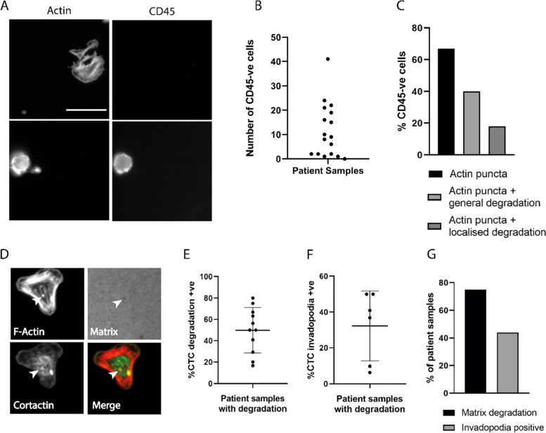
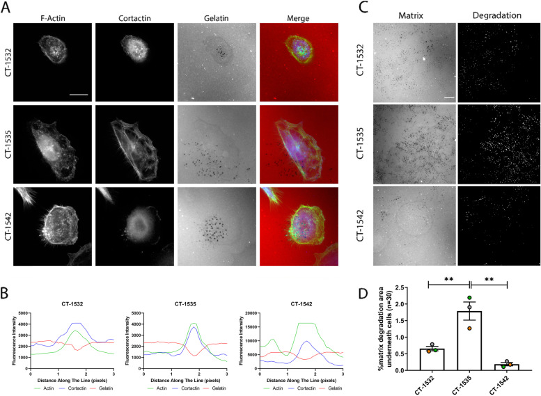
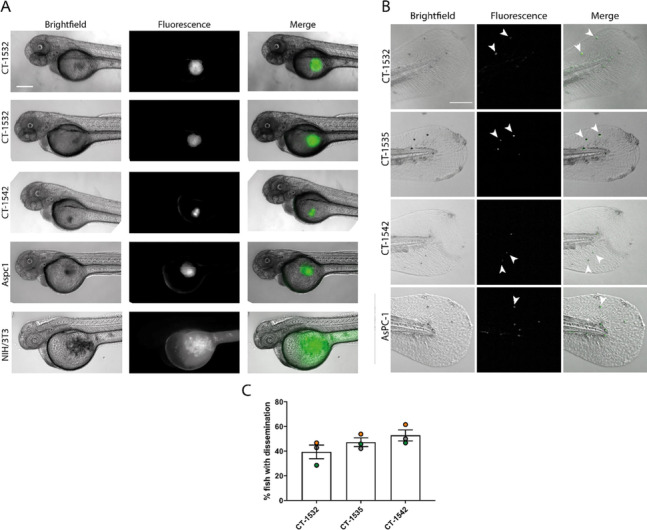
Retention of E-cadherin and N-cadherin expression indicated a transitional state of EMT progression, consistent with the idea of partial EMT that has been frequently observed in aggressive prostate cancer. All cell lines tested were capable of spontaneous invadopodia formation and possess a significant degradative ability in-vitro under basal conditions. These cell lines were invasive in-vivo and produced visible metastasis in the zebrafish dissemination assay. Importantly we have proceeded to demonstrate that circulating tumour cells isolated from prostate cancer patients exhibit invadopodia-like structures and degrade matrix with visible puncta. This work supports a role for invadopodia activity as one of the mechanisms of dissemination employed by prostate cancer cells.
The combination of studies presented here provide clear evidence that invadopodia activity can play a role in prostate cancer progression.
侵袭伪足是富含肌动蛋白的结构,在与细胞外基质的界面处以点状方式释放金属蛋白酶,被认为是肿瘤侵袭的重要驱动因素。在许多来源于多种肿瘤类型的转移性细胞系中,已经在体外和体内观察到侵袭伪足的形成。然而,前列腺癌细胞系通常不会被报道形成侵袭伪足,而且少数情况下总是需要外部刺激。
在这项研究中,从未完全表征过的原发性前列腺腺癌细胞系的体外侵袭伪足测定和体内斑马鱼传播测定中,研究了其侵袭潜力。随后,从前列腺癌患者中分离循环肿瘤细胞,并在侵袭伪足测定中进行测试。
E-钙黏蛋白和 N-钙黏蛋白表达的保留表明 EMT 进展的过渡状态,这与在侵袭性前列腺癌中经常观察到的部分 EMT 观点一致。所有测试的细胞系都能够自发形成侵袭伪足,并且在基础条件下具有显著的体外降解能力。这些细胞系在体内具有侵袭性,并在斑马鱼传播测定中产生可见的转移。重要的是,我们已经证明从前列腺癌患者中分离的循环肿瘤细胞表现出侵袭伪足样结构,并通过可见的斑点降解基质。这项工作支持侵袭伪足活性作为前列腺癌细胞扩散机制之一的作用。
这里呈现的研究组合提供了明确的证据,表明侵袭伪足活性可以在前列腺癌进展中发挥作用。