• 文献检索
  • 文档翻译
  • 深度研究
  • 学术资讯
  • Suppr Zotero 插件Zotero 插件
  • 邀请有礼
  • 套餐&价格
  • 历史记录
应用&插件
Suppr Zotero 插件Zotero 插件浏览器插件Mac 客户端Windows 客户端微信小程序
定价
高级版会员购买积分包购买API积分包
服务
文献检索文档翻译深度研究API 文档MCP 服务
关于我们
关于 Suppr公司介绍联系我们用户协议隐私条款
关注我们

Suppr 超能文献

核心技术专利:CN118964589B侵权必究
粤ICP备2023148730 号-1Suppr @ 2026

文献检索

告别复杂PubMed语法,用中文像聊天一样搜索,搜遍4000万医学文献。AI智能推荐,让科研检索更轻松。

立即免费搜索

文件翻译

保留排版,准确专业,支持PDF/Word/PPT等文件格式,支持 12+语言互译。

免费翻译文档

深度研究

AI帮你快速写综述,25分钟生成高质量综述,智能提取关键信息,辅助科研写作。

立即免费体验

扩展的CUG重复RNA在强直性肌营养不良模型细胞中诱导早衰。

Expanded CUG Repeat RNA Induces Premature Senescence in Myotonic Dystrophy Model Cells.

作者信息

Hasuike Yuhei, Mochizuki Hideki, Nakamori Masayuki

机构信息

Department of Neurology, Osaka University Graduate School of Medicine, Osaka, Japan.

出版信息

Front Genet. 2022 Mar 25;13:865811. doi: 10.3389/fgene.2022.865811. eCollection 2022.

DOI:10.3389/fgene.2022.865811
PMID:35401669
原文链接:https://pmc.ncbi.nlm.nih.gov/articles/PMC8990169/
Abstract

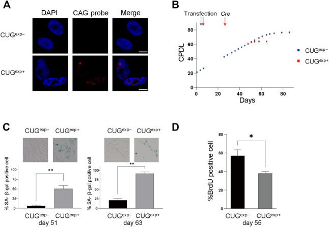
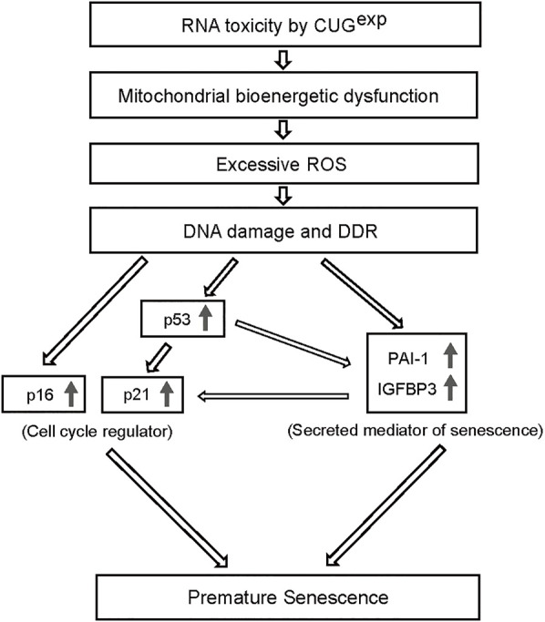
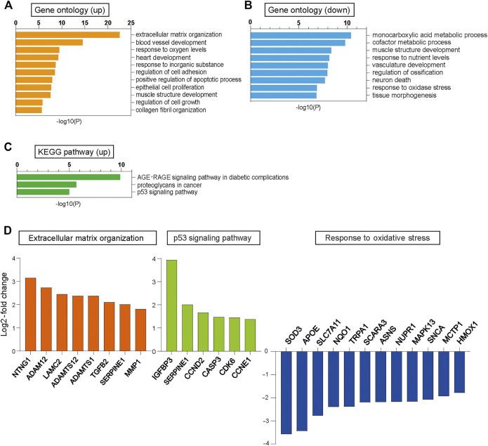
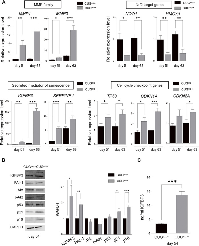
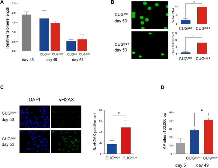
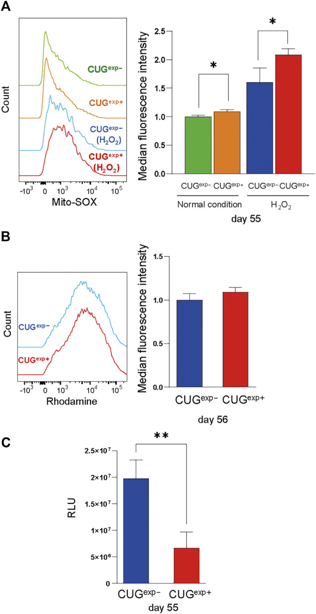
Myotonic dystrophy type 1 (DM1) is a dominantly inherited disorder due to a toxic gain of function of RNA transcripts containing expanded CUG repeats (CUG). Patients with DM1 present with multisystemic symptoms, such as muscle wasting, cognitive impairment, cataract, frontal baldness, and endocrine defects, which resemble accelerated aging. Although the involvement of cellular senescence, a critical component of aging, was suggested in studies of DM1 patient-derived cells, the detailed mechanism of cellular senescence caused by CUG RNA remains unelucidated. Here, we developed a DM1 cell model that conditionally expressed CUG RNA in human primary cells so that we could perform a detailed assessment that eliminated the variability in primary cells from different origins. Our DM1 model cells demonstrated that CUG RNA expression induced cellular senescence by a telomere-independent mechanism. Furthermore, the toxic RNA expression caused mitochondrial dysfunction, excessive reactive oxygen species production, and DNA damage and response, resulting in the senescence-associated increase of cell cycle inhibitors p21 and p16 and secreted mediators insulin-like growth factor binding protein 3 (IGFBP3) and plasminogen activator inhibitor-1 (PAI-1). This study provides unequivocal evidence of the induction of premature senescence by CUG RNA in our DM1 model cells.

摘要

1型强直性肌营养不良症(DM1)是一种常染色体显性遗传性疾病,由含有扩展CUG重复序列(CUG)的RNA转录本功能获得性毒性所致。DM1患者表现出多系统症状,如肌肉萎缩、认知障碍、白内障、前额秃发和内分泌缺陷,这些症状类似于加速衰老。尽管在对DM1患者来源细胞的研究中提示细胞衰老(衰老的一个关键组成部分)参与其中,但由CUG RNA导致细胞衰老的详细机制仍不清楚。在此,我们构建了一种DM1细胞模型,该模型可在人原代细胞中条件性表达CUG RNA,以便我们能够进行详细评估,消除不同来源原代细胞的变异性。我们的DM1模型细胞表明,CUG RNA表达通过一种不依赖端粒的机制诱导细胞衰老。此外,毒性RNA表达导致线粒体功能障碍、过量活性氧生成以及DNA损伤和反应,从而导致细胞周期抑制剂p21和p16以及分泌介质胰岛素样生长因子结合蛋白3(IGFBP3)和纤溶酶原激活物抑制剂-1(PAI-1)与衰老相关的增加。本研究为我们的DM1模型细胞中CUG RNA诱导早衰提供了明确证据。

https://cdn.ncbi.nlm.nih.gov/pmc/blobs/b1cc/8990169/223c657311a4/fgene-13-865811-g006.jpg
https://cdn.ncbi.nlm.nih.gov/pmc/blobs/b1cc/8990169/cb407f345546/fgene-13-865811-g001.jpg
https://cdn.ncbi.nlm.nih.gov/pmc/blobs/b1cc/8990169/b36336e3ddf9/fgene-13-865811-g002.jpg
https://cdn.ncbi.nlm.nih.gov/pmc/blobs/b1cc/8990169/cf7c266b13f4/fgene-13-865811-g003.jpg
https://cdn.ncbi.nlm.nih.gov/pmc/blobs/b1cc/8990169/f968eabba716/fgene-13-865811-g004.jpg
https://cdn.ncbi.nlm.nih.gov/pmc/blobs/b1cc/8990169/6149a636319a/fgene-13-865811-g005.jpg
https://cdn.ncbi.nlm.nih.gov/pmc/blobs/b1cc/8990169/223c657311a4/fgene-13-865811-g006.jpg

相似文献

1
Expanded CUG Repeat RNA Induces Premature Senescence in Myotonic Dystrophy Model Cells.扩展的CUG重复RNA在强直性肌营养不良模型细胞中诱导早衰。
Front Genet. 2022 Mar 25;13:865811. doi: 10.3389/fgene.2022.865811. eCollection 2022.
2
Cellular Senescence and Aging in Myotonic Dystrophy.肌肉萎缩性侧索硬化症中的细胞衰老和老化。
Int J Mol Sci. 2022 Feb 20;23(4):2339. doi: 10.3390/ijms23042339.
3
Short antisense-locked nucleic acids (all-LNAs) correct alternative splicing abnormalities in myotonic dystrophy.短反义锁核酸(全锁核酸)可纠正强直性肌营养不良中的异常剪接。
Nucleic Acids Res. 2015 Mar 31;43(6):3318-31. doi: 10.1093/nar/gkv163. Epub 2015 Mar 9.
4
MBNL1 and CUGBP1 modify expanded CUG-induced toxicity in a Drosophila model of myotonic dystrophy type 1.MBNL1和CUGBP1在1型强直性肌营养不良果蝇模型中调节扩展CUG诱导的毒性。
Hum Mol Genet. 2006 Jul 1;15(13):2138-45. doi: 10.1093/hmg/ddl137. Epub 2006 May 24.
5
Rationally designed small molecules that target both the DNA and RNA causing myotonic dystrophy type 1.针对导致 1 型肌强直性营养不良的 DNA 和 RNA 的合理设计的小分子。
J Am Chem Soc. 2015 Nov 11;137(44):14180-9. doi: 10.1021/jacs.5b09266. Epub 2015 Nov 3.
6
A novel CUG(exp)·MBNL1 inhibitor with therapeutic potential for myotonic dystrophy type 1.一种新型的 CUG(exp)·MBNL1 抑制剂,具有治疗 1 型肌强直性营养不良的潜力。
ACS Chem Biol. 2013 May 17;8(5):1037-43. doi: 10.1021/cb400046u. Epub 2013 Mar 20.
7
Precise small-molecule cleavage of an r(CUG) repeat expansion in a myotonic dystrophy mouse model.精准切割肌强直性营养不良小鼠模型中的 r(CUG)重复扩展的小分子。
Proc Natl Acad Sci U S A. 2019 Apr 16;116(16):7799-7804. doi: 10.1073/pnas.1901484116. Epub 2019 Mar 29.
8
RNA interference targeting CUG repeats in a mouse model of myotonic dystrophy.靶向肌强直性营养不良小鼠模型中 CUG 重复的 RNA 干扰。
Mol Ther. 2013 Feb;21(2):380-7. doi: 10.1038/mt.2012.222. Epub 2012 Nov 27.
9
A CTG repeat-selective chemical screen identifies microtubule inhibitors as selective modulators of toxic CUG RNA levels.一种 CTG 重复序列选择性化学筛选方法,可识别微管抑制剂作为毒性 CUG RNA 水平的选择性调节剂。
Proc Natl Acad Sci U S A. 2019 Oct 15;116(42):20991-21000. doi: 10.1073/pnas.1901893116. Epub 2019 Sep 30.
10
Daunorubicin reduces MBNL1 sequestration caused by CUG-repeat expansion and rescues cardiac dysfunctions in a model of myotonic dystrophy.柔红霉素减少 CUG 重复扩展引起的 MBNL1 隔离,并挽救肌强直性营养不良模型中的心脏功能障碍。
Dis Model Mech. 2018 Apr 23;11(4):dmm032557. doi: 10.1242/dmm.032557.

引用本文的文献

1
Molecular genetics of myotonic dystrophy and the evolution of therapeutic approaches.强直性肌营养不良的分子遗传学与治疗方法的演变
J Hum Genet. 2025 Jul 3. doi: 10.1038/s10038-025-01358-6.
2
Coenzyme Q improves mitochondrial and muscle dysfunction caused by CUG expanded repeats in Caenorhabditis elegans.辅酶Q可改善秀丽隐杆线虫中由CUG重复序列扩增引起的线粒体和肌肉功能障碍。
Genetics. 2025 Feb 5;229(2). doi: 10.1093/genetics/iyae208.
3
New Horizons in Myotonic Dystrophy Type 1: Cellular Senescence as a Therapeutic Target.1型强直性肌营养不良的新视野:细胞衰老作为治疗靶点

本文引用的文献

1
Cellular Senescence and Aging in Myotonic Dystrophy.肌肉萎缩性侧索硬化症中的细胞衰老和老化。
Int J Mol Sci. 2022 Feb 20;23(4):2339. doi: 10.3390/ijms23042339.
2
Transcriptome Analysis in a Primary Human Muscle Cell Differentiation Model for Myotonic Dystrophy Type 1.肌强直性营养不良 1 型的原发性人肌细胞分化模型中的转录组分析。
Int J Mol Sci. 2021 Aug 10;22(16):8607. doi: 10.3390/ijms22168607.
3
Population-Based Prevalence of Myotonic Dystrophy Type 1 Using Genetic Analysis of Statewide Blood Screening Program.基于全州血液筛查计划的遗传分析的 1 型肌强直性营养不良的人群患病率。
Bioessays. 2025 Mar;47(3):e202400216. doi: 10.1002/bies.202400216. Epub 2024 Dec 26.
4
A Novel Class of FKBP12 Ligands Rescues Premature Aging Phenotypes Associated with Myotonic Dystrophy Type 1.一类新型FKBP12配体可挽救与1型强直性肌营养不良相关的早衰表型。
Cells. 2024 Nov 22;13(23):1939. doi: 10.3390/cells13231939.
5
Fetal Brain MRI Findings in Myotonic Dystrophy and Considerations for Prenatal Genetic Testing.强直性肌营养不良的胎儿脑磁共振成像表现及产前基因检测的考量
Neurol Genet. 2024 Oct 4;10(6):e200171. doi: 10.1212/NXG.0000000000200171. eCollection 2024 Dec.
6
Common Characteristics Between Frailty and Myotonic Dystrophy Type 1: A Narrative Review.脆弱与1型强直性肌营养不良之间的共同特征:一篇叙述性综述。
Aging Dis. 2024 Aug 29;16(4):2120-2131. doi: 10.14336/AD.2024.0950.
7
Clearance of defective muscle stem cells by senolytics restores myogenesis in myotonic dystrophy type 1.衰老细胞清除剂清除有缺陷的肌肉干细胞可恢复肌强直性营养不良 1 型的肌生成。
Nat Commun. 2023 Jul 19;14(1):4033. doi: 10.1038/s41467-023-39663-3.
8
Individual transcriptomic response to strength training for patients with myotonic dystrophy type 1.1 型肌强直性营养不良患者抗阻训练的个体转录组反应。
JCI Insight. 2023 Jul 24;8(14):e163856. doi: 10.1172/jci.insight.163856.
Neurology. 2021 Feb 16;96(7):e1045-e1053. doi: 10.1212/WNL.0000000000011425. Epub 2021 Jan 20.
4
Transcriptome alterations in myotonic dystrophy frontal cortex.强直性肌营养不良症额叶皮质的转录组改变。
Cell Rep. 2021 Jan 19;34(3):108634. doi: 10.1016/j.celrep.2020.108634.
5
Relationship Between Mitochondrial Structure and Bioenergetics in Dermal Fibroblasts.真皮成纤维细胞中线粒体结构与生物能量学的关系
Front Cell Dev Biol. 2020 Dec 17;8:610266. doi: 10.3389/fcell.2020.610266. eCollection 2020.
6
Cellular senescence in ageing: from mechanisms to therapeutic opportunities.细胞衰老与衰老:从机制到治疗机会。
Nat Rev Mol Cell Biol. 2021 Feb;22(2):75-95. doi: 10.1038/s41580-020-00314-w. Epub 2020 Dec 16.
7
The hallmarks of myotonic dystrophy type 1 muscle dysfunction.1型强直性肌营养不良症肌肉功能障碍的特征
Biol Rev Camb Philos Soc. 2021 Apr;96(2):716-730. doi: 10.1111/brv.12674. Epub 2020 Dec 2.
8
PAI-1, the Plasminogen System, and Skeletal Muscle.纤溶酶原激活物抑制物-1、纤溶酶原系统与骨骼肌
Int J Mol Sci. 2020 Sep 25;21(19):7066. doi: 10.3390/ijms21197066.
9
Myotonic Dystrophy type 1 cells display impaired metabolism and mitochondrial dysfunction that are reversed by metformin.1 型肌强直性营养不良细胞表现出代谢受损和线粒体功能障碍,二甲双胍可逆转这些异常。
Aging (Albany NY). 2020 Apr 8;12(7):6260-6275. doi: 10.18632/aging.103022.
10
Role of p53 in the Regulation of Cellular Senescence.p53 在细胞衰老调控中的作用。
Biomolecules. 2020 Mar 8;10(3):420. doi: 10.3390/biom10030420.