Paolucci Iwan, Sandu Raluca-Maria, Sahli Luca, Prevost Gian Andrea, Storni Federico, Candinas Daniel, Weber Stefan, Lachenmayer Anja
ARTORG Center for Biomedical Engineering ResearchUniversity of Bern Bern Switzerland.
Department of Visceral Surgery and Medicine, Inselspital, Bern University HospitalUniversity of Bern Bern Switzerland.
IEEE Open J Eng Med Biol. 2020 Feb 14;1:3-8. doi: 10.1109/OJEMB.2019.2961094. eCollection 2020.
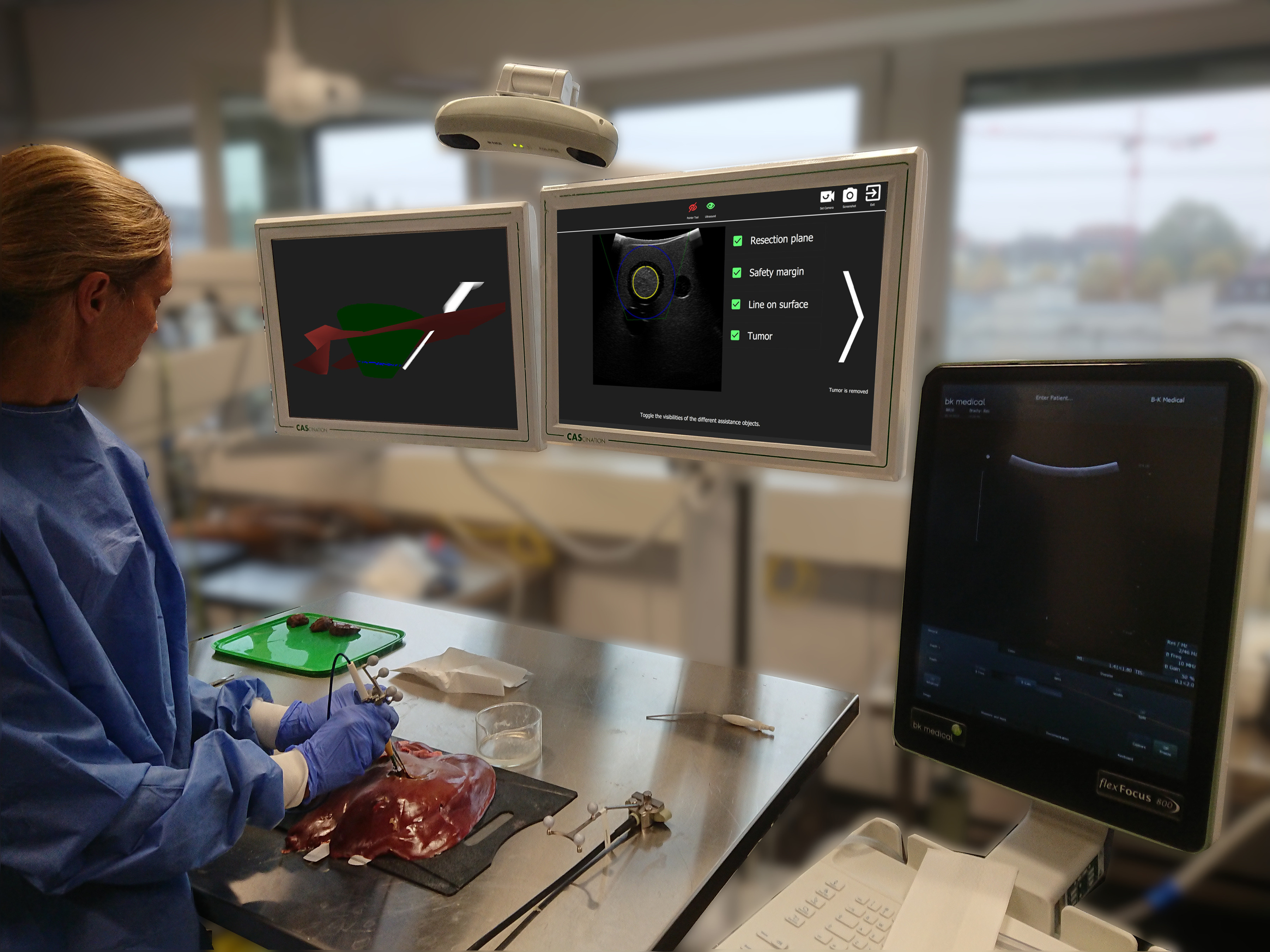
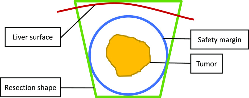
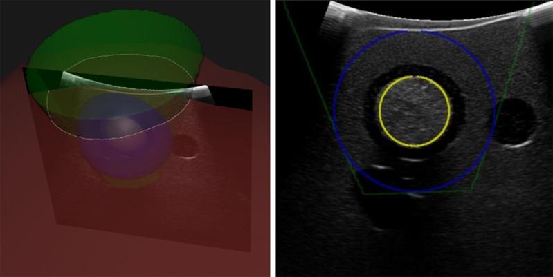
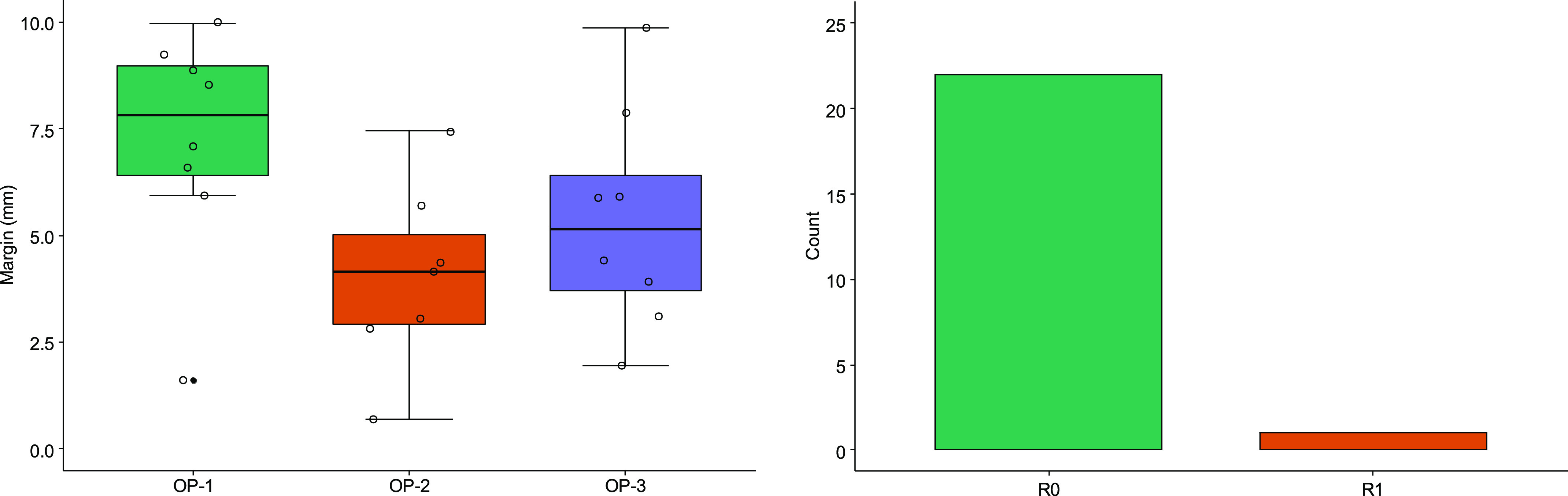
Non-anatomical resections of liver tumors can be very challenging as the surgeon cannot use anatomical landmarks on the liver surface or in the ultrasound image for guidance. This makes it difficult to achieve negative resection margins (R0) and still preserve as much healthy liver tissue as possible. Even though image-guided surgery systems have been introduced to overcome this challenge, they are still rarely used due to their inaccuracy, time-effort and complexity in usage and setup. We have developed a novel approach, which allows us to create an intra-operative resection plan using navigated ultrasound. First, the surface is scanned using a navigated ultrasound, followed by tumor segmentation on a midsection ultrasound image. Based on this information, the navigation system calculates an optimal resection strategy and displays it along with the tracked surgical instruments. In this study, this approach was evaluated by three experienced hepatobiliary surgeons on ex-vivo porcine models. Using this technique, an R0 resection could be achieved in 22 out of 23 (95.7% R0 resection rate) cases with a median resection margin of 5.9 mm (IQR 3.5-7.7 mm). The resection margin between operators 1, 2 and 3 was 7.8 mm, 4.15 mm and 5.1 mm respectively (p = 0.054). This approach could represent a useful tool for intra-operative guidance in non-anatomical resection alongside conventional ultrasound guidance. However, instructions and training are essential especially if the operator has not used an image-guidance system before.
肝脏肿瘤的非解剖性切除极具挑战性,因为外科医生无法利用肝脏表面或超声图像中的解剖标志进行引导。这使得实现阴性切缘(R0)并尽可能保留更多健康肝组织变得困难。尽管已引入图像引导手术系统来克服这一挑战,但由于其准确性、使用和设置的时间精力以及复杂性,它们仍然很少被使用。我们开发了一种新方法,该方法使我们能够使用导航超声创建术中切除计划。首先,使用导航超声扫描肝脏表面,然后在肝脏中部超声图像上进行肿瘤分割。基于这些信息,导航系统计算出最佳切除策略,并将其与跟踪的手术器械一起显示出来。在本研究中,三位经验丰富的肝胆外科医生在离体猪模型上对该方法进行了评估。使用该技术,23例中有22例(R0切除率为95.7%)实现了R0切除,中位切缘为5.9毫米(四分位间距为3.5 - 7.7毫米)。操作者1、2和3的切缘分别为7.8毫米、4.15毫米和5.1毫米(p = 0.054)。这种方法可能是一种在非解剖性切除术中与传统超声引导并行的有用的术中引导工具。然而,特别是如果操作者以前未使用过图像引导系统,那么说明和培训至关重要。