Kumar Ankur, Kala Niranjan, Telles Shirley
Division of Yoga and Clinical Neurophysiology, Patanjali Research Foundation, Patanjali Yogpeeth, Haridwar, Uttarakhand, India.
Int J Yoga. 2022 Jan-Apr;15(1):19-24. doi: 10.4103/ijoy.ijoy_179_21. Epub 2022 Mar 21.
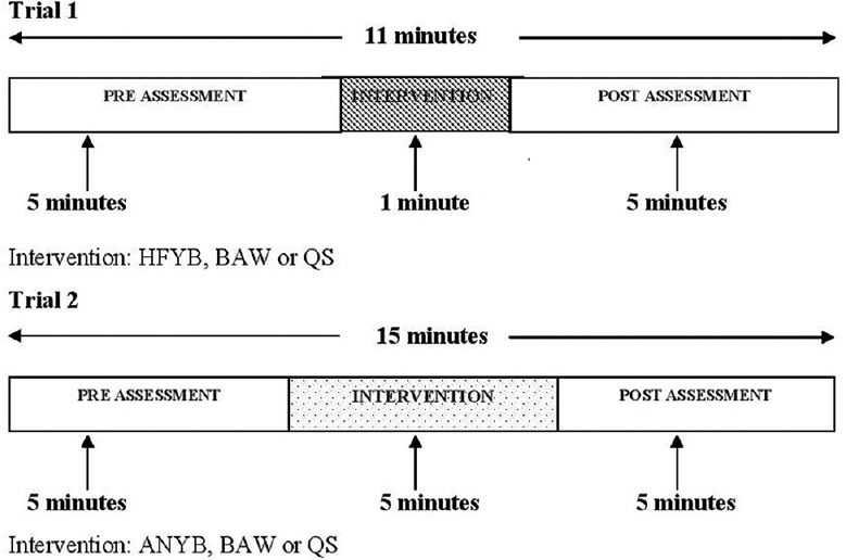
Breath frequency can alter cerebral blood flow. The study aimed to determine bilateral middle cerebral arterial hemodynamics in high-frequency yoga breathing (HFYB) and slow frequency alternate nostril yoga breathing (ANYB) using transcranial Doppler sonography.
Healthy male volunteers were assessed in two separate trials before, during, and after HFYB (2.0 Hz for 1 min, = 16) and ANYB (12 breaths per minute for 5 min, = 22). HFYB and ANYB were separately compared to breath awareness (BAW) and to control sessions.
The data were analyzed using repeated-measures ANOVA with Bonferroni adjusted tests.
During HFYB there was a decrease in end-diastolic velocity (EDV) and mean flow velocity (MFV) ( < 0.01 for left and < 0.05 for right middle cerebral arteries; MCA) with an increase in pulsatility index (PI) for the right MCA ( < 0.05). During ANYB, there was a bilateral decrease in peak systolic velocity ( < 0.05 for left and < 0.01 for right MCA), EDV ( < 0.01) and MFV ( < 0.01 for left and < 0.001 for right MCA) and an increase in PI ( < 0.01). During BAW of the two sessions there was a decrease in lateralized flow and end-diastolic velocities ( < 0.05) and an increase in PI ( < 0.05).
Changes in peak flow velocities and pulsatility indices during and after HFYB, ANYB, and BAW suggest decreased cerebrovascular blood flow and increased flow resistance based on different mechanisms.
呼吸频率可改变脑血流量。本研究旨在使用经颅多普勒超声测定高频瑜伽呼吸(HFYB)和低频交替鼻孔瑜伽呼吸(ANYB)时双侧大脑中动脉的血流动力学。
在两项单独试验中,对健康男性志愿者在进行HFYB(2.0赫兹,持续1分钟,n = 16)和ANYB(每分钟12次呼吸,持续5分钟,n = 22)之前、期间和之后进行评估。分别将HFYB和ANYB与呼吸觉知(BAW)及对照时段进行比较。
采用重复测量方差分析及Bonferroni校正t检验对数据进行分析。
在HFYB期间,舒张末期速度(EDV)和平均流速(MFV)降低(左侧大脑中动脉P < 0.01,右侧大脑中动脉P < 0.05),右侧大脑中动脉搏动指数(PI)增加(P < 0.05)。在ANYB期间,双侧收缩期峰值速度降低(左侧大脑中动脉P < 0.05,右侧大脑中动脉P < 0.01)、EDV降低(P < 0.01)和MFV降低(左侧大脑中动脉P < 0.01,右侧大脑中动脉P < 0.001),PI增加(P < 0.01)。在两个时段的BAW期间,侧化血流和舒张末期速度降低(P < 0.05),PI增加(P < 0.05)。
HFYB、ANYB和BAW期间及之后峰值流速和搏动指数的变化表明,基于不同机制,脑血管血流量减少且血流阻力增加。