Chen Senlin, Wang Ying, Wu Xianyong, Chang Jianchao, Jin Weiming, Li Wei, Song Peiwen, Wu Yuanyuan, Zhu Jiajia, Qian Yinfeng, Shen Cailiang, Yu Yongqiang, Dong Fulong
Department of Orthopedics, Department of Spine Surgery, The First Affiliated Hospital of AnHui Medical University, Hefei, China.
Department of Radiology, The First Affiliated Hospital of AnHui Medical University, Hefei, China.
Front Aging Neurosci. 2022 Apr 4;14:784263. doi: 10.3389/fnagi.2022.784263. eCollection 2022.
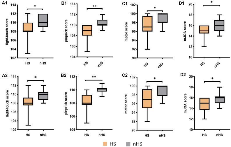
Degenerative cervical myelopathy is a progressive neurodegenerative disease, that has become increasingly prevalent in the aging population worldwide. The current study determined the factors affecting degeneration in the sensorimotor tract with degenerative cervical myelopathy and its relationship with brain structure. We divided patients into hyperintensity (HS) and non-hyperintensity (nHS) groups and measured the fractional anisotropy and apparent diffusion coefficients of the lateral corticospinal tract (CST), fasciculus gracilis and fasciculus cuneatus (FGC). Voxel-based morphometry (VBM) and tract-based spatial statistics (TBSS) techniques were used to estimate brain structure changes. Correlation of the modified Japanese Orthopaedic Association (mJOA) score, light touch, pinprick, motor score, and fractional anisotropy (FA) ratios of the CST at different levels were analyzed. Compared to healthy controls, the FA ratios of CST in the HS and nHS groups were decreased at all levels, and the apparent diffusion coefficient (ADC) ratio was increased only at C4/5 levels in the HS group. The FA ratio of FGC was decreased at the C3/4 and C4/5 levels in the HS group and only decreased at the C4/5 level in the nHS group. The ADC ratio was decreased only at the C4/5 level in the HS group. VBM analysis revealed that the volume of the precentral gyrus, postcentral gyrus, and paracentral lobule increased in patients compared to controls. TBSS analysis found no statistical significance between the sensory and motor tracts in white matter. The volume of clusters in HS and nHS groups negatively correlated with the C1/2 FA ratio of the CST. The results showed that the degeneration distance of the CST was longer than the FGC, and the degeneration distance was related to the degree of compression and spinal cord damage. Structural compensation and the neurotrophin family may lead to enlargement of the brain.
退行性颈椎脊髓病是一种进行性神经退行性疾病,在全球老龄化人口中日益普遍。本研究确定了影响退行性颈椎脊髓病感觉运动束退变的因素及其与脑结构的关系。我们将患者分为高信号(HS)组和非高信号(nHS)组,测量了皮质脊髓侧束(CST)、薄束和楔束(FGC)的各向异性分数和表观扩散系数。采用基于体素的形态学测量(VBM)和基于束的空间统计学(TBSS)技术来评估脑结构变化。分析了改良日本骨科学会(mJOA)评分、轻触觉、针刺觉、运动评分以及不同节段CST的各向异性分数(FA)比值之间的相关性。与健康对照组相比,HS组和nHS组各节段CST的FA比值均降低,HS组仅在C4/5节段表观扩散系数(ADC)比值升高。HS组FGC在C3/4和C4/5节段的FA比值降低,nHS组仅在C4/5节段降低。HS组仅在C4/5节段ADC比值降低。VBM分析显示,与对照组相比,患者中央前回、中央后回和中央旁小叶体积增大。TBSS分析发现白质感觉束和运动束之间无统计学差异。HS组和nHS组簇体积与CST的C1/2 FA比值呈负相关。结果表明,CST的退变距离长于FGC,退变距离与压迫程度和脊髓损伤程度有关。结构代偿和神经营养因子家族可能导致脑体积增大。