Upper Gastrointestinal Translational Research Laboratory, Peter MacCallum Cancer Centre, Melbourne, Australia.
The Sir Peter MacCallum Department of Oncology, The University of Melbourne, Melbourne, Australia.
BMC Gastroenterol. 2022 Apr 21;22(1):197. doi: 10.1186/s12876-022-02268-z.
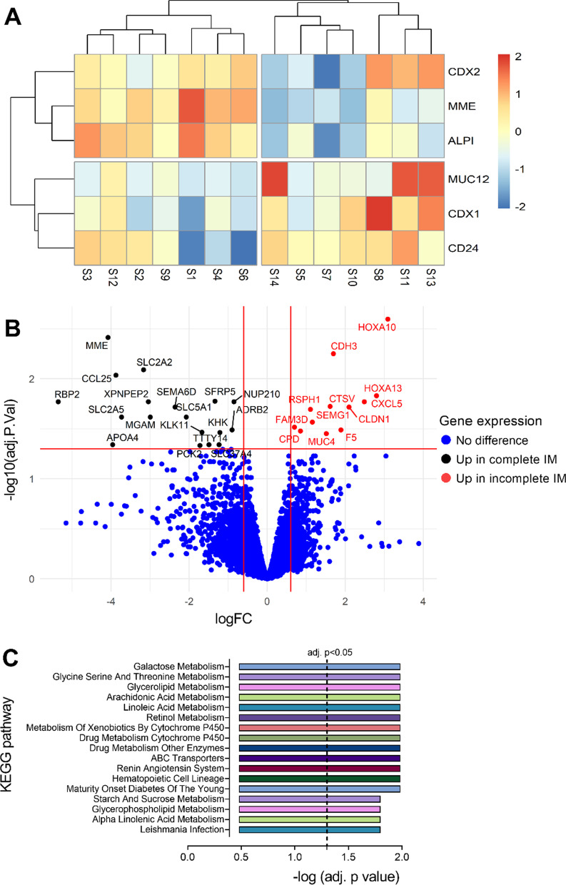
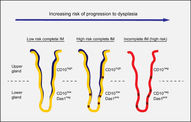
Intestinal metaplasia (IM) is considered a key pivot point in the Correa model of gastric cancer (GC). It is histologically subtyped into the complete and incomplete subtypes, the latter being associated with a greater risk of progression. However, the clinical utility of IM subtyping remains unclear, partially due to the absence of reliable defining biomarkers.
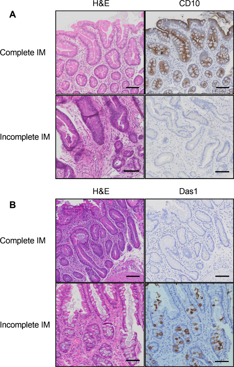
Based on gene expression data and existing literature, we selected CD10 and Das1 as candidate biomarkers to distinguish complete and incomplete IM glands in tissues from patients without GC (IM-GC) and patients with GC (IM + GC). Immunohistochemical staining of individually subtyped IM glands was scored after blinding by two researchers using tissue belonging to both IM-GC and IM + GC patients. Whole tissue Das1 staining was further assessed using digital image quantification (cellSens Dimension, Olympus).
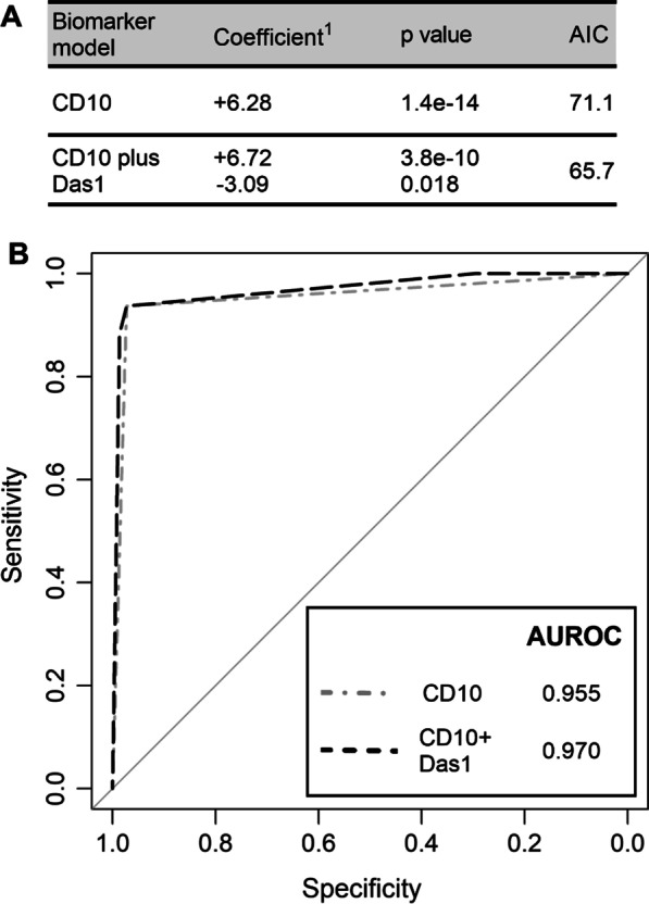
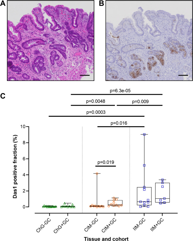
Across both cohorts CD10 stained the IM brush border and was shown to have a high sensitivity (87.5% and 94.9% in IM-GC and IM + GC patients respectively) and specificity (100.0% and 96.7% respectively) with an overall AUROC of 0.944 for complete IM glands. By contrast Das1 stained mainly goblet cells and the apical membrane of epithelial cells, mostly of incomplete IM glands with a low sensitivity (28.6% and 29.3% in IM-GC and IM + GC patients respectively) but high specificity (98.3% and 85.1% respectively) and an overall AUROC of 0.603 for incomplete IM glands. A combined logistic regression model showed a significant increase in AUROC for detecting complete IM glands (0.955 vs 0.970). Whole tissue digital quantification of Das1 staining showed a significant association with incomplete IM compared to complete IM, both in IM-GC and in IM + GC patients (p = 0.016 and p = 0.009 respectively, Mann-Whitney test and unpaired t test used). Additionally, complete IM in IM + GC patients exhibited significantly more Das1 staining than in IM-GC patients (p = 0.019, Mann-Whitney test).
These findings suggest that CD10 is an outstanding biomarker for complete IM and Das1 may be useful as a secondary biomarker for IM glands at greater risk of progression irrespective of IM subtype. Overall, the clinical use of these biomarkers could lead to improved patient stratification and targeted surveillance.
肠上皮化生(IM)被认为是胃癌(GC)科雷亚模型中的关键转折点。它在组织学上分为完全型和不完全型,后者与更大的进展风险相关。然而,IM 分型的临床实用性仍不清楚,部分原因是缺乏可靠的定义生物标志物。
基于基因表达数据和现有文献,我们选择 CD10 和 Das1 作为候选生物标志物,以区分无 GC(IM-GC)和有 GC(IM+GC)患者组织中的完全和不完全 IM 腺体。两名研究人员在对组织进行盲法评分后,对单独分型的 IM 腺体进行免疫组织化学染色,这些组织来自 IM-GC 和 IM+GC 患者。使用数字图像定量(Olympus 的 cellSens Dimension)进一步评估整个组织的 Das1 染色。
在两个队列中,CD10 均染色 IM 刷状缘,具有高灵敏度(分别为 IM-GC 和 IM+GC 患者的 87.5%和 94.9%)和特异性(分别为 100.0%和 96.7%),总 AUROC 为 0.944,用于完全型 IM 腺体。相比之下,Das1 主要染色杯状细胞和上皮细胞的顶膜,主要是不完全型 IM 腺体,灵敏度低(分别为 IM-GC 和 IM+GC 患者的 28.6%和 29.3%),但特异性高(分别为 98.3%和 85.1%),总 AUROC 为 0.603,用于不完全型 IM 腺体。联合逻辑回归模型显示,检测完全型 IM 腺体的 AUROC 显著增加(0.955 对 0.970)。Das1 染色的全组织数字定量显示,与完全型 IM 相比,在 IM-GC 和 IM+GC 患者中,与不完全型 IM 有显著关联(p=0.016 和 p=0.009,分别为 Mann-Whitney 检验和未配对 t 检验)。此外,与 IM-GC 患者相比,IM+GC 患者的完全型 IM 中 Das1 染色明显更多(p=0.019,Mann-Whitney 检验)。
这些发现表明,CD10 是完全型 IM 的出色生物标志物,而 Das1 可能是一种有用的生物标志物,用于检测无论 IM 亚型如何,进展风险更高的 IM 腺体。总体而言,这些生物标志物的临床应用可能会导致患者分层和靶向监测的改善。