• 文献检索
  • 文档翻译
  • 深度研究
  • 学术资讯
  • Suppr Zotero 插件Zotero 插件
  • 邀请有礼
  • 套餐&价格
  • 历史记录
应用&插件
Suppr Zotero 插件Zotero 插件浏览器插件Mac 客户端Windows 客户端微信小程序
定价
高级版会员购买积分包购买API积分包
服务
文献检索文档翻译深度研究API 文档MCP 服务
关于我们
关于 Suppr公司介绍联系我们用户协议隐私条款
关注我们

Suppr 超能文献

核心技术专利:CN118964589B侵权必究
粤ICP备2023148730 号-1Suppr @ 2026

文献检索

告别复杂PubMed语法,用中文像聊天一样搜索,搜遍4000万医学文献。AI智能推荐,让科研检索更轻松。

立即免费搜索

文件翻译

保留排版,准确专业,支持PDF/Word/PPT等文件格式,支持 12+语言互译。

免费翻译文档

深度研究

AI帮你快速写综述,25分钟生成高质量综述,智能提取关键信息,辅助科研写作。

立即免费体验

颌面部骨良性间充质牙源性肿瘤:3例报告及3年随访

Benign mesenchymal odontogenic tumors of the maxillofacial bones: A report of 3 cases with 3 years follow-up.

作者信息

Berberi Antoine N, Aoun Georges T, Aad Georges P, Khalaf Emile N

机构信息

Department of Oral and Maxillofacial Surgery, Faculty of Dental Medicine, Lebanese University, Beirut, Lebanon.

Oral Medicine and Maxillofacial Radiology, Faculty of Dental Medicine, Lebanese University, Beirut, Lebanon.

出版信息

J Oral Maxillofac Pathol. 2022 Feb;26(Suppl 1):S46-S50. doi: 10.4103/jomfp.jomfp_94_21. Epub 2022 Feb 28.

DOI:10.4103/jomfp.jomfp_94_21
PMID:35450255
原文链接:https://pmc.ncbi.nlm.nih.gov/articles/PMC9017847/
Abstract

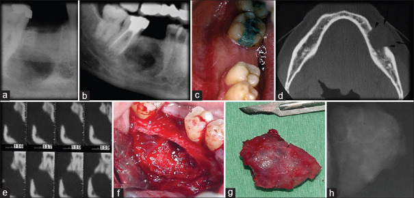
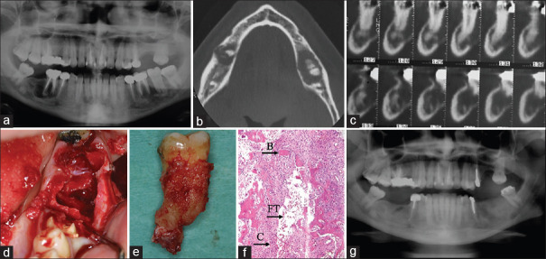
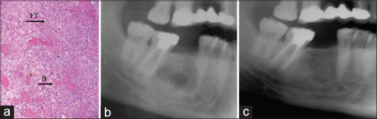
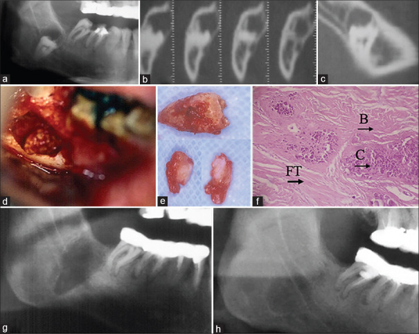
Benign mesenchymal odontogenic tumors are lesions derived from the mesenchymal components of the tooth-forming apparatus and are consequently found within the jawbone. Benign fibro-osseous tumors are part of this category of lesions in which normal bone is substituted, initially by fibrous tissue and within time become infiltrated by osteoid and cementoid elements. They are asymptomatic, slow-growing lesions and remain undiagnosed until swelling of the face becomes prominent and they share similar radiological characteristics. Herein, we report three cases of ossifying fibroma, cemento-osseous fibroma and periapical cemento-osseous dysplasia and analyze all the correlating factors, clinical history, radiological and histological features, intraoperative appearance, and treatment with a 3-year follow-up period. Despite the advances in the identification of these pathologies, clinicians still face difficulties in their classification and the diagnosis due to overlap in both histological and radiographic findings. An accurate final diagnosis is essential for appropriate treatment and an informative prognosis.

摘要

良性间充质牙源性肿瘤是源自牙齿形成器官间充质成分的病变,因此见于颌骨内。良性纤维-骨肿瘤是这类病变的一部分,其中正常骨最初被纤维组织替代,随着时间推移被类骨质和牙骨质成分浸润。它们是无症状、生长缓慢的病变,直到面部肿胀明显才被诊断出来,并且它们具有相似的放射学特征。在此,我们报告3例骨化性纤维瘤、牙骨质-骨纤维瘤和根尖周牙骨质-骨发育异常病例,并分析所有相关因素、临床病史、放射学和组织学特征、术中表现以及3年随访期的治疗情况。尽管在这些病变的识别方面取得了进展,但由于组织学和影像学表现存在重叠,临床医生在其分类和诊断方面仍面临困难。准确的最终诊断对于恰当的治疗和提供信息的预后至关重要。

https://cdn.ncbi.nlm.nih.gov/pmc/blobs/e47b/9017847/49b59b2d0990/JOMFP-26-46-g004.jpg
https://cdn.ncbi.nlm.nih.gov/pmc/blobs/e47b/9017847/40889fc6d89e/JOMFP-26-46-g001.jpg
https://cdn.ncbi.nlm.nih.gov/pmc/blobs/e47b/9017847/b563642b5d92/JOMFP-26-46-g002.jpg
https://cdn.ncbi.nlm.nih.gov/pmc/blobs/e47b/9017847/49e501a82500/JOMFP-26-46-g003.jpg
https://cdn.ncbi.nlm.nih.gov/pmc/blobs/e47b/9017847/49b59b2d0990/JOMFP-26-46-g004.jpg

相似文献

1
Benign mesenchymal odontogenic tumors of the maxillofacial bones: A report of 3 cases with 3 years follow-up.颌面部骨良性间充质牙源性肿瘤:3例报告及3年随访
J Oral Maxillofac Pathol. 2022 Feb;26(Suppl 1):S46-S50. doi: 10.4103/jomfp.jomfp_94_21. Epub 2022 Feb 28.
2
A Rare Case of Cemento-Ossifying Fibroma: A Case Report.骨化性纤维瘤罕见病例:一例报告
Cureus. 2023 May 7;15(5):e38685. doi: 10.7759/cureus.38685. eCollection 2023 May.
3
The importance of clinical and radiological findings for the definitive histopathologic diagnosis of benign fibro-osseous lesions of the jaws: Study of 276 cases.临床及影像学表现对颌骨良性纤维-骨病变确诊性组织病理学诊断的重要性:276例病例研究
J Stomatol Oral Maxillofac Surg. 2022 Jun;123(3):364-371. doi: 10.1016/j.jormas.2021.04.008. Epub 2021 Apr 18.
4
Imaging in the diagnosis of cemento-ossifying fibroma: a case series.骨化性纤维瘤诊断中的影像学:病例系列
J Clin Imaging Sci. 2012;2:52. doi: 10.4103/2156-7514.100373. Epub 2012 Aug 30.
5
Can non-collagenous proteins be employed for the differential diagnosis among fibrous dysplasia, cemento-osseous dysplasia and cemento-ossifying fibroma?非胶原蛋白能否用于纤维结构不良、骨化性骨纤维发育不良和骨化性纤维瘤的鉴别诊断?
Pathol Res Pract. 2019 Jul;215(7):152450. doi: 10.1016/j.prp.2019.152450. Epub 2019 May 13.
6
Ossifying Fibroma of Non-odontogenic Origin: A Fibro-osseous Lesion in the Craniofacial Skeleton to be (Re-)considered.非牙源性骨化性纤维瘤:一种需(重新)审视的颅面骨骼纤维-骨病变
Head Neck Pathol. 2022 Mar;16(1):257-267. doi: 10.1007/s12105-021-01351-3. Epub 2021 Jun 26.
7
Distinguishing features of focal cemento-osseous dysplasias and cemento-ossifying fibromas: I. A pathologic spectrum of 316 cases.局灶性牙骨质-骨发育异常和牙骨质化纤维瘤的鉴别特征:I. 316例病例的病理谱
Oral Surg Oral Med Oral Pathol Oral Radiol Endod. 1997 Sep;84(3):301-9. doi: 10.1016/s1079-2104(97)90348-6.
8
Cemento-Ossifying Fibroma in Maxillofacial Region: A Series of 16 Cases.颌面部骨化性纤维瘤:16例病例系列报道
J Maxillofac Oral Surg. 2021 Jun;20(2):240-245. doi: 10.1007/s12663-019-01304-y. Epub 2019 Nov 8.
9
Central ossifying fibroma, periapical cemento-osseous dysplasia and complex odon-toma occurring in the same jaw.发生于同一颌骨的中央性骨化性纤维瘤、根尖周牙骨质骨发育异常和复合性牙瘤。
Clin Pract. 2011 May 17;1(2):e36. doi: 10.4081/cp.2011.e36. eCollection 2011 May 16.
10
A case report of cemento-ossifying fibroma.骨化性纤维瘤病例报告
J Maxillofac Oral Surg. 2010 Jun;9(2):178-81. doi: 10.1007/s12663-010-0061-4. Epub 2010 Sep 22.

本文引用的文献

1
Clinical assessment of cemento-osseous dysplasia based on three-dimensional diagnostic imaging: A case report.基于三维诊断成像的骨-牙骨质发育不良的临床评估:病例报告。
Aust Endod J. 2021 Apr;47(1):105-112. doi: 10.1111/aej.12488. Epub 2021 Feb 1.
2
Classification and nomenclature of fibro-osseous lesions.
Oral Surg Oral Med Oral Pathol Oral Radiol. 2021 Apr;131(4):385-389. doi: 10.1016/j.oooo.2020.12.004. Epub 2021 Jan 29.
3
Difficulties in the diagnosis of periapical translucencies and in the classification of cemento-osseous dysplasia.根尖透光区诊断困难及骨-牙骨质发育不良分类困难。
BMC Oral Health. 2019 Jul 10;19(1):139. doi: 10.1186/s12903-019-0843-0.
4
Benign Fibro-Osseous Lesions of the Head and Neck.头颈部良性纤维-骨病变
Head Neck Pathol. 2019 Sep;13(3):466-475. doi: 10.1007/s12105-018-0992-5. Epub 2019 Mar 18.
5
Cemento-Osseous Dysplasias: Imaging Features Based on Cone Beam Computed Tomography Scans.骨水泥发育异常:基于锥形束计算机断层扫描的影像特征
Braz Dent J. 2018 Jan-Feb;29(1):99-104. doi: 10.1590/0103-6440201801621.
6
Cemento-ossifying fibroma of the mandible. A clinicopathological report.下颌骨骨化性纤维瘤。一份临床病理报告。
Saudi Med J. 2017 May;38(5):541-545. doi: 10.15537/smj.2017.5.15643.
7
Update from the 4th Edition of the World Health Organization Classification of Head and Neck Tumours: Odontogenic and Maxillofacial Bone Tumors.世界卫生组织头颈部肿瘤分类第4版更新:牙源性和颌面骨肿瘤
Head Neck Pathol. 2017 Mar;11(1):68-77. doi: 10.1007/s12105-017-0794-1. Epub 2017 Feb 28.
8
Diagnosis and management of benign fibro-osseous lesions of the jaws: a current review for the dental clinician.颌骨良性纤维-骨病变的诊断与管理:牙科临床医生的最新综述
Oral Dis. 2017 May;23(4):440-450. doi: 10.1111/odi.12531. Epub 2016 Aug 1.
9
Cemento-ossifying fibroma of the jaw.颌骨骨化性纤维瘤
BMJ Case Rep. 2016 May 12;2016:bcr2015214327. doi: 10.1136/bcr-2015-214327.
10
Two cases of multiple ossifying fibromas in the jaws.两例颌骨多发性骨化性纤维瘤。
Diagn Pathol. 2014 Mar 28;9:75. doi: 10.1186/1746-1596-9-75.