Kim Hye Jin, Gong Eun Jeong, Bang Chang Seok, Lee Jae Jun, Suk Ki Tae, Baik Gwang Ho
Department of Internal Medicine, Hallym University College of Medicine, Chuncheon 24253, Korea.
Institute for Liver and Digestive Diseases, Hallym University, Chuncheon 24253, Korea.
J Pers Med. 2022 Apr 17;12(4):644. doi: 10.3390/jpm12040644.
Wireless capsule endoscopy allows the identification of small intestinal protruded lesions, such as polyps, tumors, or venous structures. However, reading wireless capsule endoscopy images or movies is time-consuming, and minute lesions are easy to miss. Computer-aided diagnosis (CAD) has been applied to improve the efficacy of the reading process of wireless capsule endoscopy images or movies. However, there are no studies that systematically determine the performance of CAD models in diagnosing gastrointestinal protruded lesions.
The aim of this study was to evaluate the diagnostic performance of CAD models for gastrointestinal protruded lesions using wireless capsule endoscopic images.
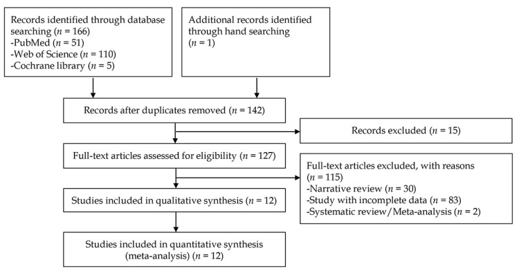
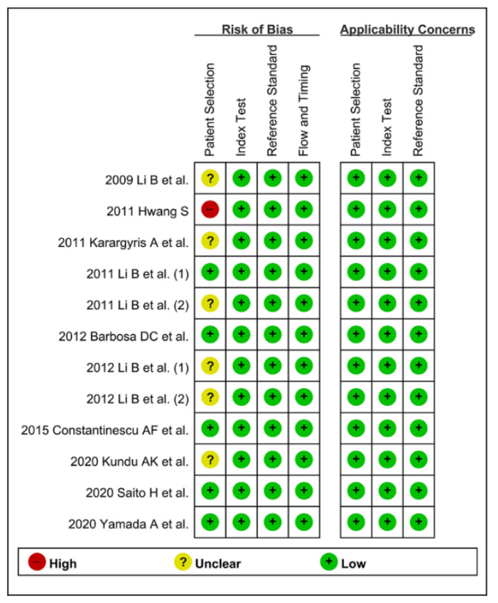
Core databases were searched for studies based on CAD models for the diagnosis of gastrointestinal protruded lesions using wireless capsule endoscopy, and data on diagnostic performance were presented. A systematic review and diagnostic test accuracy meta-analysis were performed.
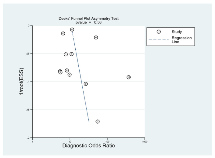
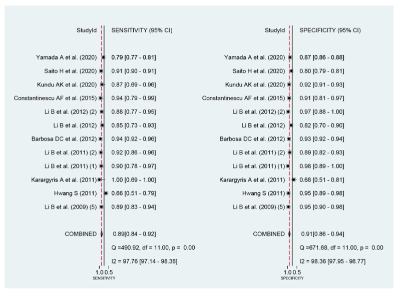
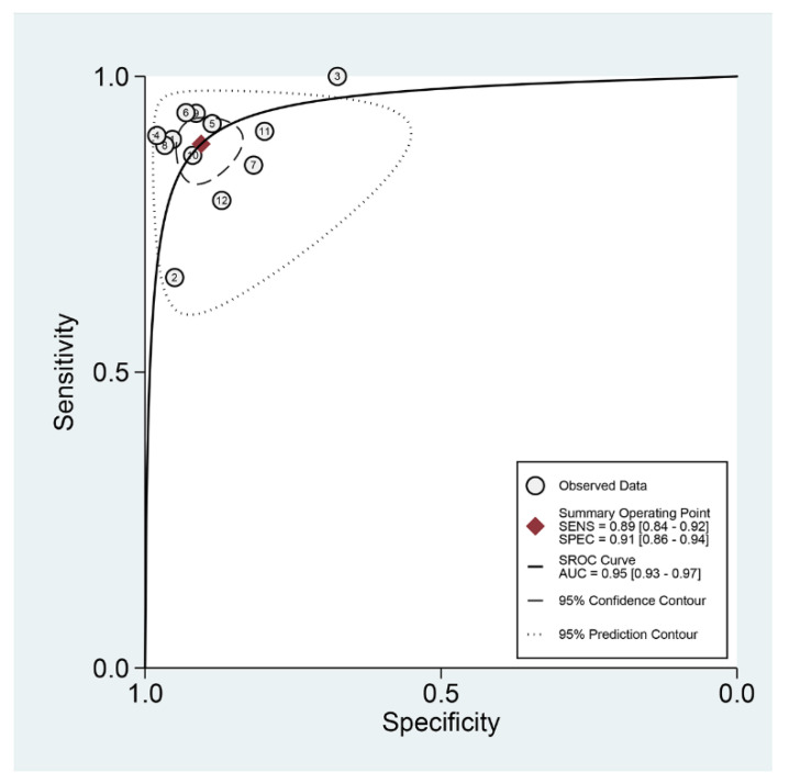
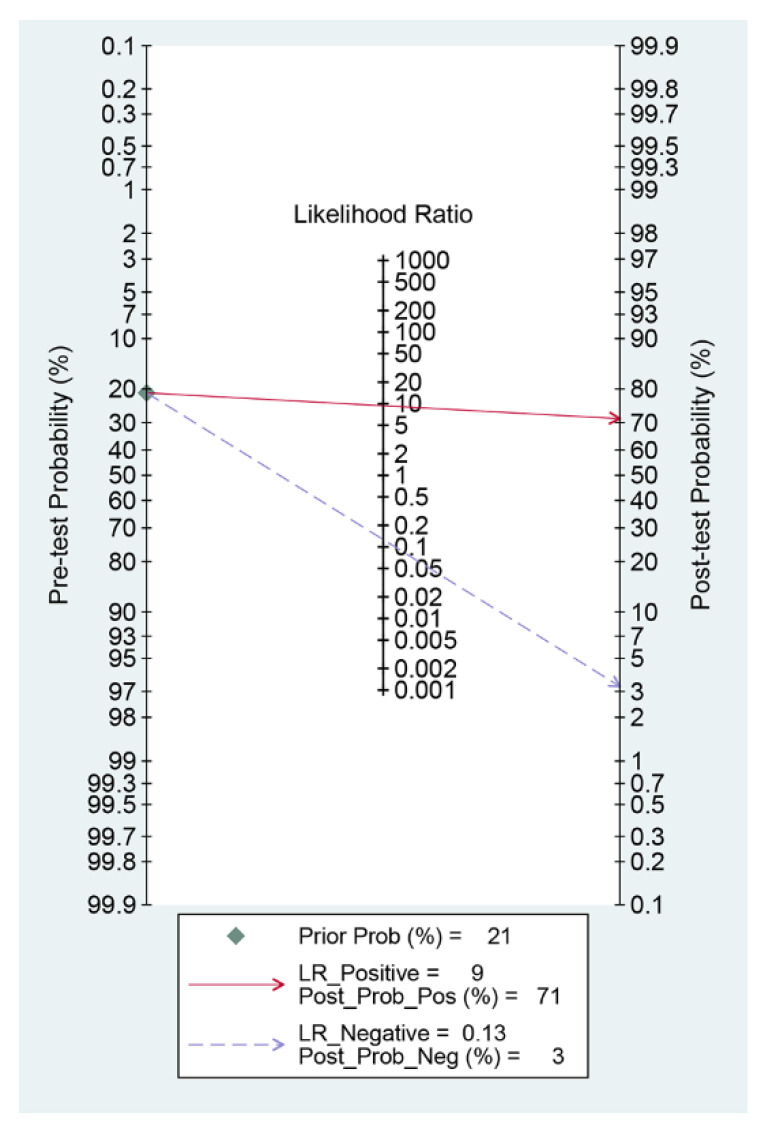
Twelve studies were included. The pooled area under the curve, sensitivity, specificity, and diagnostic odds ratio of CAD models for the diagnosis of protruded lesions were 0.95 (95% confidence interval, 0.93-0.97), 0.89 (0.84-0.92), 0.91 (0.86-0.94), and 74 (43-126), respectively. Subgroup analyses showed robust results. Meta-regression found no source of heterogeneity. Publication bias was not detected.
CAD models showed high performance for the optical diagnosis of gastrointestinal protruded lesions based on wireless capsule endoscopy.
无线胶囊内镜可用于识别小肠突出性病变,如息肉、肿瘤或静脉结构。然而,阅读无线胶囊内镜图像或视频很耗时,微小病变容易漏诊。计算机辅助诊断(CAD)已被应用于提高无线胶囊内镜图像或视频阅读过程的效率。然而,尚无系统评估CAD模型诊断胃肠道突出性病变性能的研究。
本研究旨在评估使用无线胶囊内镜图像的CAD模型对胃肠道突出性病变的诊断性能。
检索核心数据库中基于CAD模型利用无线胶囊内镜诊断胃肠道突出性病变的研究,并呈现诊断性能数据。进行系统评价和诊断试验准确性的Meta分析。
纳入12项研究。CAD模型诊断突出性病变的合并曲线下面积、敏感性、特异性和诊断比值比分别为0.95(95%置信区间,0.93 - 0.97)、0.89(0.84 - 0.92)、0.91(0.86 - 0.94)和74(43 - 126)。亚组分析结果稳健。Meta回归未发现异质性来源。未检测到发表偏倚。
基于无线胶囊内镜,CAD模型在胃肠道突出性病变的光学诊断中表现出高性能。