Brugger Marcus, Laschinger Melanie, Lampl Sandra, Schneider Annika, Manske Katrin, Esfandyari Dena, Hüser Norbert, Hartmann Daniel, Steiger Katja, Engelhardt Stefan, Wohlleber Dirk, Knolle Percy A
Institute of Molecular Immunology and Experimental Oncology, School of Medicine, Technical University of Munich, Germany.
Department of Internal Medicine I, School of Medicine, University Hospital München rechts der Isar, Technical University of Munich, Germany.
JHEP Rep. 2022 Mar 6;4(5):100465. doi: 10.1016/j.jhepr.2022.100465. eCollection 2022 May.
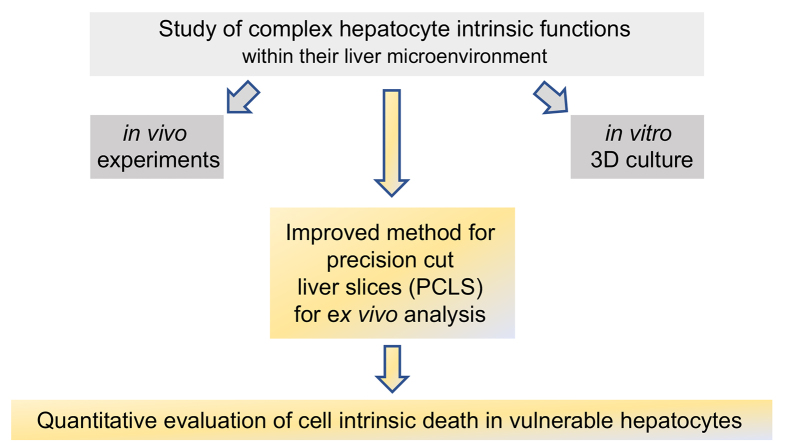
BACKGROUND & AIMS: Increased sensitivity towards tumor necrosis factor (TNF)-induced cell death in virus-infected hepatocytes has revealed a so far unrecognized hepatocyte-intrinsic antiviral immune surveillance mechanism, for which no or model is available. We aimed to establish precision-cut liver slices (PCLS) as a model system to study hepatocyte-intrinsic regulation of apoptosis.
Preparation of PCLS from mouse and human liver tissue was optimized for minimal procedure-associated apoptosis. Functionality of liver cells in PCLS was characterized using extracellular flux analysis to determine mitochondrial respiration, and viral infection with recombinant adenovirus and lymphocytic choriomeningitis virus (LCMV) was used to probe for hepatocyte-intrinsic sensitivity towards apoptosis in PCLS. Apoptosis was detected by immunohistochemical staining for cleaved-caspase 3 and quantified by detection of effector caspase activity in PCLS.
We established an optimized protocol for preparation of PCLS from human and mouse models using agarose-embedding of liver tissue to improve precision cutting and using organ-protective buffer solutions to minimize procedure-associated cell death. PCLS prepared from virus-infected livers showed preserved functional metabolic properties. Importantly, in PCLS from adenovirus- and LCMV-infected livers we detected increased induction of apoptosis after TNF challenge .
We conclude that PCLS can be used as model system to characterize hepatocyte-intrinsic sensitivity to cell death. This may also enable researchers to characterize human hepatocyte sensitivity to apoptosis in PCLS prepared from patients with acute or chronic liver diseases.
Virus-infected hepatocytes show an increased sensitivity towards induction of cell death signaling through the TNF receptor. Studying this hepatocyte-intrinsic antiviral immune surveillance mechanism has been hampered by the absence of model systems that reciprocate the finding of increased apoptosis of virus-infected hepatocytes challenged with TNF. Herein, we report that an optimized protocol for generation of precision-cut liver slices can be used to study this hepatocyte-intrinsic surveillance mechanism .
病毒感染的肝细胞对肿瘤坏死因子(TNF)诱导的细胞死亡敏感性增加,揭示了一种迄今未被认识的肝细胞固有抗病毒免疫监视机制,目前尚无相关模型。我们旨在建立精密肝切片(PCLS)作为研究肝细胞固有凋亡调控的模型系统。
优化从小鼠和人类肝脏组织制备PCLS的方法,以尽量减少与操作相关的凋亡。使用细胞外通量分析来确定线粒体呼吸,以表征PCLS中肝细胞的功能,并用重组腺病毒和淋巴细胞性脉络丛脑膜炎病毒(LCMV)感染来探究PCLS中肝细胞对凋亡的固有敏感性。通过免疫组化染色检测裂解的半胱天冬酶-3来检测凋亡,并通过检测PCLS中的效应半胱天冬酶活性进行定量。
我们建立了一种优化方案,用于从人和小鼠模型制备PCLS,采用肝组织琼脂糖包埋以提高切割精度,并使用器官保护缓冲液以尽量减少与操作相关的细胞死亡。从病毒感染的肝脏制备的PCLS显示出保留的功能代谢特性。重要的是,在来自腺病毒和LCMV感染肝脏的PCLS中,我们检测到TNF刺激后凋亡诱导增加。
我们得出结论,PCLS可作为模型系统来表征肝细胞对细胞死亡的固有敏感性。这也可能使研究人员能够表征从急性或慢性肝病患者制备的PCLS中人类肝细胞对凋亡的敏感性。
病毒感染的肝细胞对通过TNF受体诱导细胞死亡信号的敏感性增加。由于缺乏能够重现TNF刺激的病毒感染肝细胞凋亡增加这一发现的模型系统,对这种肝细胞固有抗病毒免疫监视机制的研究受到了阻碍。在此,我们报告一种优化的精密肝切片生成方案可用于研究这种肝细胞固有监视机制。