Hu Xiaoxin, Li Jianwen, Sun Yinan, Sun Yiqun, Tong Tong
Department of Radiology, Fudan University Shanghai Cancer Center; Department of Oncology, Shanghai Medical College, Fudan University, Shanghai, China.
Department of Urology, Dushu Lake Hospital Affiliated To Soochow University, Medical Center of Soochow University, Suzhou Dushu Lake Hospital, Suzhou, China.
Front Oncol. 2022 Apr 7;12:616310. doi: 10.3389/fonc.2022.616310. eCollection 2022.
The purpose of the study was to assess the ability of percentage of tumor invasion (PTI) of T3 rectal cancer on pretreatment MRI as an imaging biomarker to reflect aggressiveness and to predict tumor response after neoadjuvant chemoradiation (NCRT) in Chinese population.
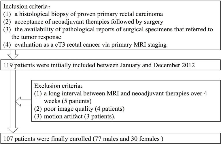
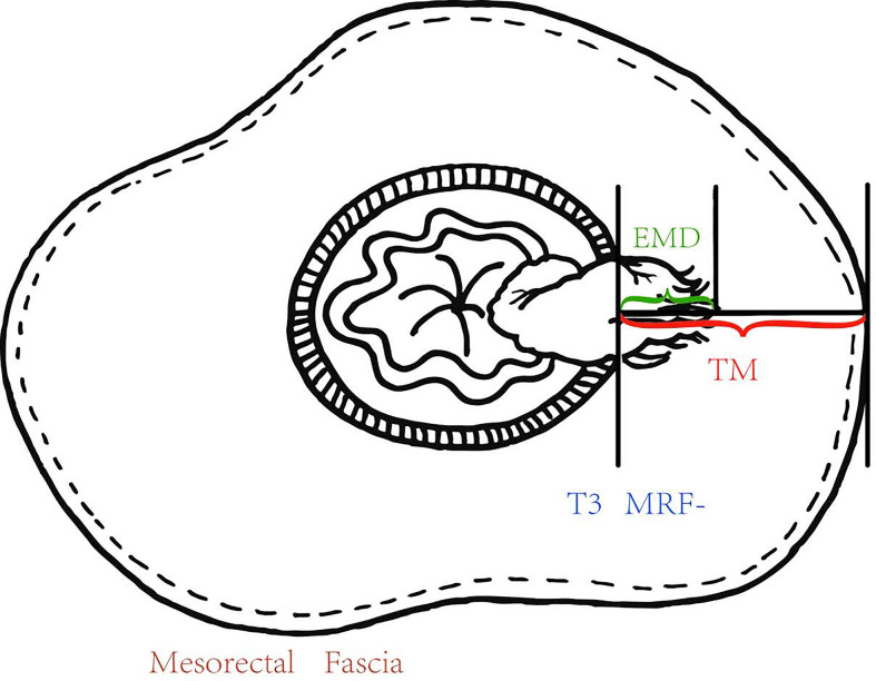
A total of 107 Chinese rectal cancer patients who underwent pretreatment MRI staging as T3 were included. The extramural depth of tumor invasion (EMD), the distance between outer border of muscularis propria (MP) and mesorectal fascia (MRF) we called "thickness of the mesorectum (TM)") at the same slice and direction were measured at pretreatment MRI, and PTI was equal to EMD/TM, was calculated. The EMD and PTI of subgroups based on pretreatment CEA, CA19-9 levels; N category and pathological complete response (pCR) were compared. The parameters, which described tumor invasion, were compared between pCR and non-pCR group. Student t-tests and logistic analysis were applied.
The pretreatment PTI was higher in CEA ≥5.2 ng/ml patients (58.52% ± 27.68%) than in CEA <5.2 ng/ml patients (47.27% ± 24.15%) ( = 0.034). The pretreatment EMD in non-pCR group (7.21 ± 2.85 mm) was higher than in pCR group (6.14 ± 3.56 mm) ( = 0.049). The pretreatment PTI in non-pCR group (57.4% ± 26.4%) was higher than in pCR group (47.3% ± 29.1%) ( = 0.041). Compared with patients with PTI ≥50%, MRF (+), more patients with PTI <50%, MRF (-) showed pCR (OR = 8.44, = 0.005; OR = 6.32, = 0.024).
The PTI obtained at pretreatment MRI may serve as an imaging biomarker to reflect tumor aggressiveness and predict which T3 rectal cancer patients may benefit from NCRT in Chinese population.
本研究旨在评估T3期直肠癌治疗前MRI的肿瘤浸润百分比(PTI)作为一种影像生物标志物反映肿瘤侵袭性以及预测中国人群新辅助放化疗(NCRT)后肿瘤反应的能力。
纳入107例经治疗前MRI分期为T3期的中国直肠癌患者。在治疗前MRI上测量肿瘤外膜浸润深度(EMD)以及同一层面和方向上固有肌层(MP)外边界与直肠系膜筋膜(MRF)之间的距离(我们称之为“直肠系膜厚度(TM)”),并计算PTI,PTI等于EMD/TM。比较基于治疗前癌胚抗原(CEA)、糖类抗原19-9(CA19-9)水平;N分期以及病理完全缓解(pCR)的亚组的EMD和PTI。比较pCR组和非pCR组之间描述肿瘤浸润的参数。采用学生t检验和逻辑分析。
CEA≥5.2 ng/ml患者的治疗前PTI(58.52%±27.68%)高于CEA<5.2 ng/ml患者(47.27%±24.15%)(P = 0.034)。非pCR组的治疗前EMD(7.21±2.85 mm)高于pCR组(6.14±3.56 mm)(P = 0.049)。非pCR组的治疗前PTI(57.4%±26.4%)高于pCR组(47.3%±29.1%)(P = 0.041)。与PTI≥50%、MRF(+)的患者相比,PTI<50%、MRF(-)的患者pCR发生率更高(比值比[OR]=8.44,P = 0.005;OR = 6.32,P = 0.024)。
治疗前MRI获得的PTI可作为一种影像生物标志物,反映肿瘤侵袭性,并预测中国人群中哪些T3期直肠癌患者可能从NCRT中获益。