Department of Neurosurgery, Gunma University Graduate School of Medicine, Maebashi, Japan.
Department of Neurosurgery, National Center Hospital, National Center of Neurology and Psychiatry, Kodaira, Japan.
Sci Rep. 2022 Apr 26;12(1):6805. doi: 10.1038/s41598-022-10753-4.
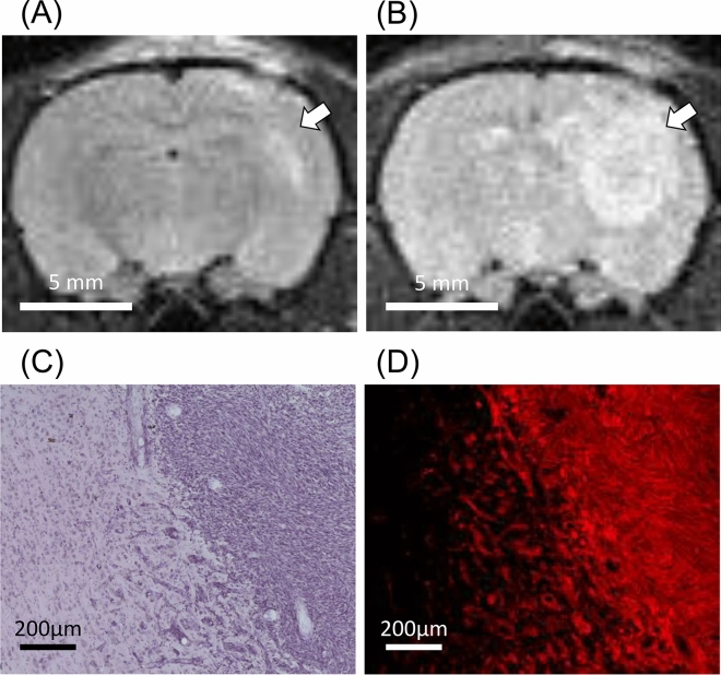
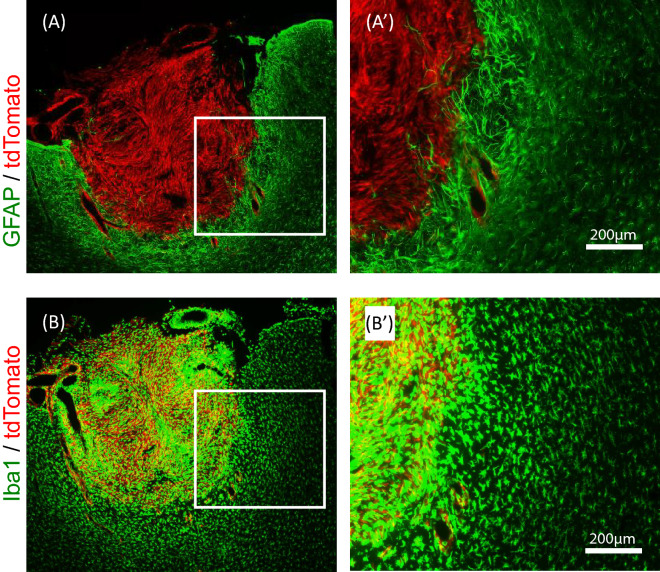
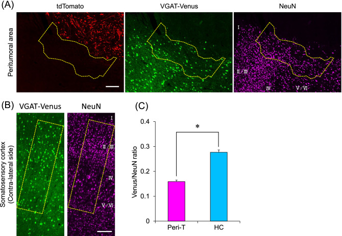
Patients with glioma often demonstrate epilepsy. We previously found burst discharges in the peritumoral area in patients with malignant brain tumors during biopsy. Therefore, we hypothesized that the peritumoral area may possess an epileptic focus and that biological alterations in the peritumoral area may cause epileptic symptoms in patients with glioma. To test our hypothesis, we developed a rat model of glioma and characterized it at the cellular and molecular levels. We first labeled rat C6 glioma cells with tdTomato, a red fluorescent protein (C6-tdTomato), and implanted them into the somatosensory cortex of VGAT-Venus rats, which specifically expressed Venus, a yellow fluorescent protein in GABAergic neurons. We observed that the density of GABAergic neurons was significantly decreased in the peritumoral area of rats with glioma compared with the contralateral healthy side. By using a combination technique of laser capture microdissection and RNA sequencing (LCM-seq) of paraformaldehyde-fixed brain sections, we demonstrated that 19 genes were differentially expressed in the peritumoral area and that five of them were associated with epilepsy and neurodevelopmental disorders. In addition, the canonical pathways actively altered in the peritumoral area were predicted to cause a reduction in GABAergic neurons. These results suggest that biological alterations in the peritumoral area may be a cause of glioma-related epilepsy.
脑胶质瘤患者常伴有癫痫。我们之前发现恶性脑肿瘤患者在活检过程中肿瘤周围区域存在爆发放电。因此,我们假设肿瘤周围区域可能存在癫痫灶,并且肿瘤周围区域的生物学改变可能导致脑胶质瘤患者出现癫痫症状。为了验证我们的假设,我们建立了脑胶质瘤大鼠模型,并在细胞和分子水平上对其进行了表征。我们首先用红色荧光蛋白(tdTomato)标记大鼠 C6 神经胶质瘤细胞(C6-tdTomato),然后将其植入特异性表达 GABA 能神经元中黄色荧光蛋白 Venus 的 VGAT-Venus 大鼠体感皮层。我们观察到与对侧健康侧相比,胶质瘤大鼠肿瘤周围区域 GABA 能神经元的密度显著降低。通过使用激光捕获显微切割和甲醛固定脑切片的 RNA 测序(LCM-seq)的组合技术,我们证明了在肿瘤周围区域有 19 个基因表达差异,其中 5 个基因与癫痫和神经发育障碍有关。此外,预测肿瘤周围区域中活跃改变的经典途径会导致 GABA 能神经元减少。这些结果表明,肿瘤周围区域的生物学改变可能是导致与脑胶质瘤相关的癫痫的原因。