Singh Kunwar I, Gollapudi Sumanth, Kumar Jyoti, Butzmann Alexandra, Small Corinn, Kreimer Sara, Saglam Emine Arzu, Warnke Roger, Silva Oscar, Ohgami Robert S
Department of Pathology, University of California, San Francisco, San Francisco, CA, United States.
Department of Pathology, Stanford University, Stanford, CA, United States.
Front Oncol. 2022 Apr 13;12:857606. doi: 10.3389/fonc.2022.857606. eCollection 2022.
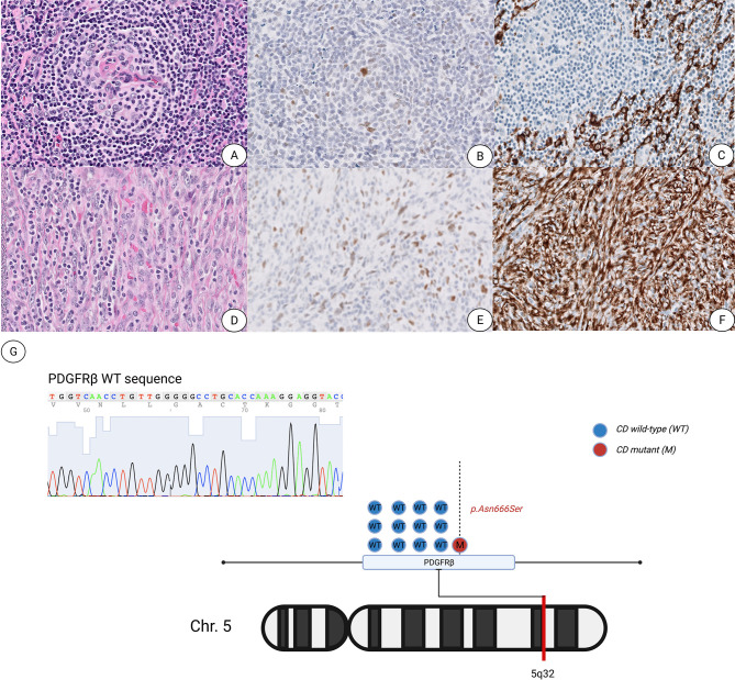
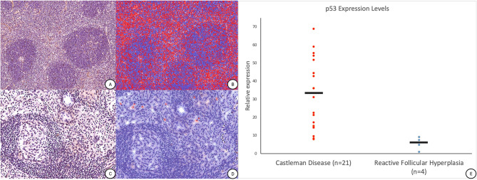
Castleman disease (CD) is a rare lymphoproliferative disorder with distinct clinical subtypes. However, our understanding of the underlying pathogenesis of particular subtypes of CD remains unclear. While the characteristic morphologic changes within UCD, including occasional cases of overgrowth of spindled stromal and follicular dendritic cells have been described, the nature and origin of these spindle cells remain elusive. Few reports have suggested that underlying stromal cells in UCD are clonally neoplastic and may be of fibroblastic reticular cell (FRC) or follicular dendritic cell (FDC) origins given their close clonal relationship. Although certain histomorphologic features may aid diagnosis, there are no specific biomarkers that can differentiate a reactive process mimicking UCD from true UCD. Hence, we describe an index case with morphology consistent with the hyaline vascular subtype of UCD with concomitant atypical smooth muscle actin (SMA)-positive stromal spindle cell proliferation containing a recurrent PDGFRB N666S mutation and upregulation of p53 expression. Further analysis of 21 additional cases of UCD identified increased p53 expression by digital image analysis and SMA positive stromal cells predominantly within the paracortical and intrafollicular areas further strengthening the hypothesis of the stromal cellular derivation and origins of UCD.
Castleman病(CD)是一种罕见的淋巴增生性疾病,具有不同的临床亚型。然而,我们对特定亚型CD的潜在发病机制仍不清楚。虽然已描述了UCD内的特征性形态学变化,包括偶尔出现的梭形基质细胞和滤泡树突状细胞过度生长的病例,但这些梭形细胞的性质和起源仍然难以捉摸。很少有报告表明,鉴于其密切的克隆关系,UCD中的潜在基质细胞是克隆性肿瘤性的,可能起源于成纤维网状细胞(FRC)或滤泡树突状细胞(FDC)。尽管某些组织形态学特征可能有助于诊断,但没有特定的生物标志物能够区分模仿UCD的反应性过程与真正的UCD。因此,我们描述了一例索引病例,其形态与UCD的透明血管亚型一致,伴有非典型平滑肌肌动蛋白(SMA)阳性的基质梭形细胞增殖,包含复发性PDGFRB N666S突变和p53表达上调。对另外21例UCD病例的进一步分析通过数字图像分析确定p53表达增加,且SMA阳性基质细胞主要位于副皮质区和滤泡内区域,进一步强化了UCD基质细胞来源和起源的假说。