Department of Pathology and Veterinary Diagnostics of the Institute of Veterinary Medicine, Warsaw University of Life Sciences, Nowoursynowska 159c, 02-776, Warsaw, Poland.
Department of Preclinical Sciences, of the Institute of Veterinary Medicine, Warsaw University of Life Sciences, Nowoursynowska 159c, 02-776, Warsaw, Poland.
BMC Vet Res. 2022 May 2;18(1):160. doi: 10.1186/s12917-022-03260-1.
To date, Campylobacter jejuni has not been found to be pathogenic to peafowl. The available publications show that out of a total of 44 samples tested from peafowl, this bacterium was isolated only in two cases. Eimeria pavonina infestations in the peafowl have been described, but no fatal cases have been reported yet.
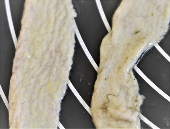
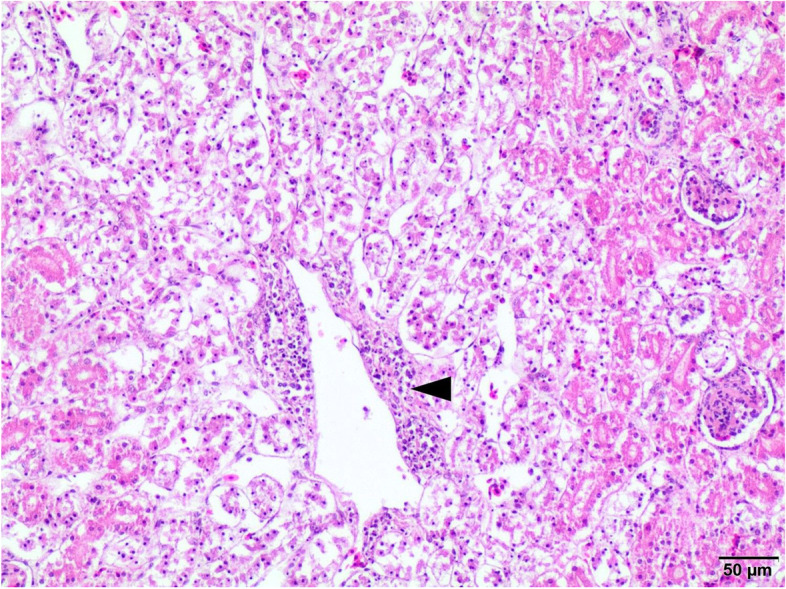
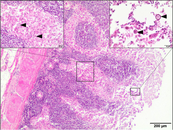
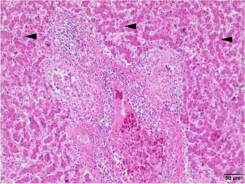
The four-year-old peacock was presented with chronic diarrhea, emaciation and weakness. Post mortem examination revealed enlarged and pale kidneys, small intestinal mucosal necrosis and thickening of intestinal wall, and pericardial effusion. The histopathological examination revealed necrotic enteritis with marked mononuclear cells infiltration associated with the presence of coccidia, additionally there was histological evidence of septicemia in liver and kidneys. Bacteria identification was based on light microscopy of the small intestine sample, culture, and biochemical tests. Further identification was based on PCR. Antimicrobial susceptibility profile was created by determination of minimal inhibitory concentration (MIC) values for 6 antimicrobial agents from 5 different classes. PCR assays were performed to detect virulence factors genes responsible for motility, cytolethal distending toxin production, adhesion and internalization. Bacteriology of the small intestine sample showed abundant growth almost exclusively of Campylobacter jejuni, resistant to ciprofloxacin, gentamycin and ampicillin. Bacteria was sensitive to Amoxicillin + clavulanic acid, tetracycline, and erythromycin. All tested virulence factors genes have been detected. The parasitological examination was performed by microscopic examination of fresh faeces and intestinal content, and revealed the moderate number of Eimeria pavonina, Histomonas meleagridis, single Capillaria spp. eggs as well Heterakis spp. like parasites.
The above case shows that a virulent isolate of Campylobacter jejuni in combination with a parasitic invasion may cause chronic enteritis in peafowl, which most likely led to extreme exhaustion of the host organism and death.
迄今为止,尚未发现空肠弯曲菌对孔雀致病。现有文献表明,在总共对 44 份孔雀样本进行的检测中,仅在两种情况下分离到这种细菌。已描述了孔雀中艾美耳球虫的感染,但尚未报告致命病例。
这只 4 岁的孔雀表现为慢性腹泻、消瘦和虚弱。剖检发现肾脏肿大且苍白、小肠黏膜坏死和肠壁增厚,以及心包积液。组织病理学检查显示伴有裂殖体的坏死性肠炎,有明显的单核细胞浸润,此外肝脏和肾脏有败血症的组织学证据。细菌鉴定基于小肠样本的光镜检查、培养和生化试验。进一步的鉴定基于 PCR。通过测定来自 5 个不同类别的 6 种抗菌药物的最小抑菌浓度(MIC)值来确定抗菌药物敏感性谱。PCR 检测用于检测与运动、细胞致死性扩张毒素产生、黏附和内化相关的毒力因子基因。小肠样本的细菌学显示几乎仅存在大量空肠弯曲菌的生长,对环丙沙星、庆大霉素和氨苄西林耐药。细菌对阿莫西林+克拉维酸、四环素和红霉素敏感。所有测试的毒力因子基因均被检出。寄生虫学检查通过新鲜粪便和肠内容物的显微镜检查进行,发现有中等数量的孔雀艾美耳球虫、火鸡组织滴虫、单个毛细线虫卵以及异刺线虫样寄生虫。
上述病例表明,强毒力空肠弯曲菌与寄生虫侵袭的组合可能导致孔雀慢性肠炎,这很可能导致宿主极度衰竭和死亡。