Zarafshan Eghbal, Rahbarghazi Reza, Rezaie Jafar, Aslani Mohammad Reza, Saberianpour Shirin, Ahmadi Mahdi, Keyhanmanesh Rana
Department of Physiology, Faculty of Medicine, Tabriz University of Medical Sciences, Tabriz, Iran.
Stem Cell Research Center, Tabriz University of Medical Sciences, Tabriz, Iran.
Adv Pharm Bull. 2022 Jan;12(1):176-182. doi: 10.34172/apb.2022.019. Epub 2020 Oct 19.
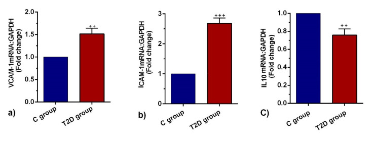
Diabetes mellitus, especially type 2, is conceived as a devastating chronic metabolic disease globally. Due to the existence of an extensive vascular network in the pulmonary tissue, it is suggested that lungs are sensitive to the diabetic condition like other tissues. This study was designed to address the possible effect of type 2 diabetes mellitus on the promotion of pathological changes via vascular injury. Sixteen male Wistar rats were randomly allocated to the two of control and T2D groups. To induce type 2 diabetes (T2D), rats were received high-fat and a single dose of streptozotocin (STZ). On week 12, rats were euthanized and lungs samples were taken. Using hematoxylin and eosin (H&E) staining, the pathological changes were monitored. The expression of intercellular adhesion molecule (ICAM-1) and vascular cell adhesion molecule 1 (VCAM-1), and interleukin 10 (IL-10) was monitored using real-time PCR assay. The level of tumor necrosis factor-α (TNF-α) was detected using ELISA assay. Nitrosative stress was monitored using the Griess assay. Pathological examination in bronchoalveolar discharge revealed the existence of mild to moderate interstitial bronchopneumonia and increased neutrophilic leukocytosis compared to the control. Enhanced ICAM-1 and VCAM-1 expression and suppression of IL-10 was found using real-time PCR analysis ( < 0.05). The levels of TNF-α and NO were increased with diabetic changes compared to the control rats ( < 0.05). T2D could promote pulmonary tissue injury via the production of TNF-α and up-regulation of vascular ICAM-1 and VCAM-1. The inflammatory status and vascular ICAM-1 and VCAM-1 increase immune cell recruitment into the pulmonary niche.
糖尿病,尤其是2型糖尿病,被认为是全球范围内一种具有毁灭性的慢性代谢疾病。由于肺组织中存在广泛的血管网络,有人提出肺和其他组织一样对糖尿病状态敏感。本研究旨在探讨2型糖尿病通过血管损伤促进病理变化的可能作用。将16只雄性Wistar大鼠随机分为对照组和T2D组。为诱导2型糖尿病(T2D),给大鼠喂食高脂饲料并注射单剂量链脲佐菌素(STZ)。在第12周,对大鼠实施安乐死并采集肺组织样本。使用苏木精和伊红(H&E)染色监测病理变化。使用实时PCR检测细胞间黏附分子(ICAM-1)、血管细胞黏附分子1(VCAM-1)和白细胞介素10(IL-10)的表达。使用ELISA检测肿瘤坏死因子-α(TNF-α)水平。使用格里斯试剂检测法监测亚硝化应激。支气管肺泡灌洗的病理检查显示,与对照组相比,存在轻度至中度间质性支气管肺炎且中性粒细胞增多。实时PCR分析发现ICAM-1和VCAM-1表达增强,IL-10受到抑制(P<0.05)。与对照大鼠相比,糖尿病改变使TNF-α和NO水平升高(P<0.05)。T2D可通过产生TNF-α以及上调血管ICAM-1和VCAM-1来促进肺组织损伤。炎症状态以及血管ICAM-1和VCAM-1增加了免疫细胞向肺微环境的募集。