Murphy Sandra, Zweyer Margit, Mundegar Rustam R, Swandulla Dieter, Ohlendieck Kay
Department of Biology, Maynooth University, National University of Ireland, Maynooth, Maynooth, Co. Kildare, Ireland.
Institute of Physiology II, University of Bonn, Bonn, D‑53115, Germany.
HRB Open Res. 2018 Sep 17;1:17. doi: 10.12688/hrbopenres.12846.2. eCollection 2018.
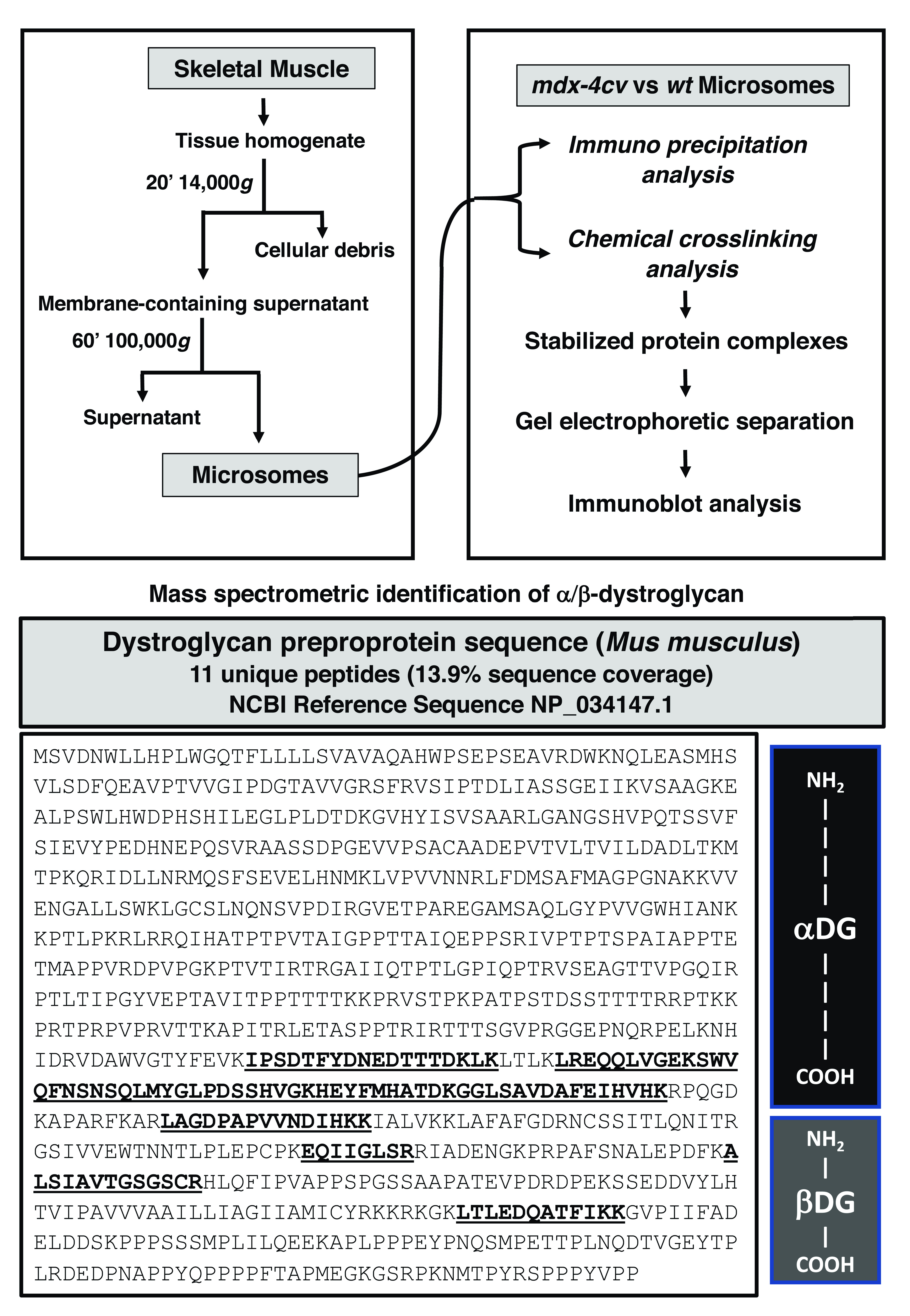
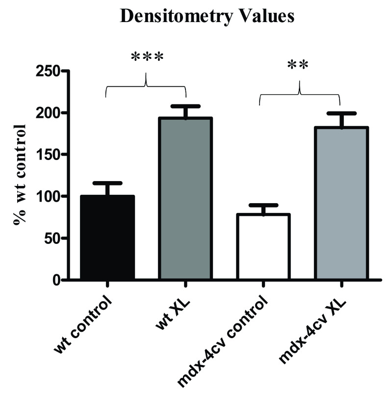
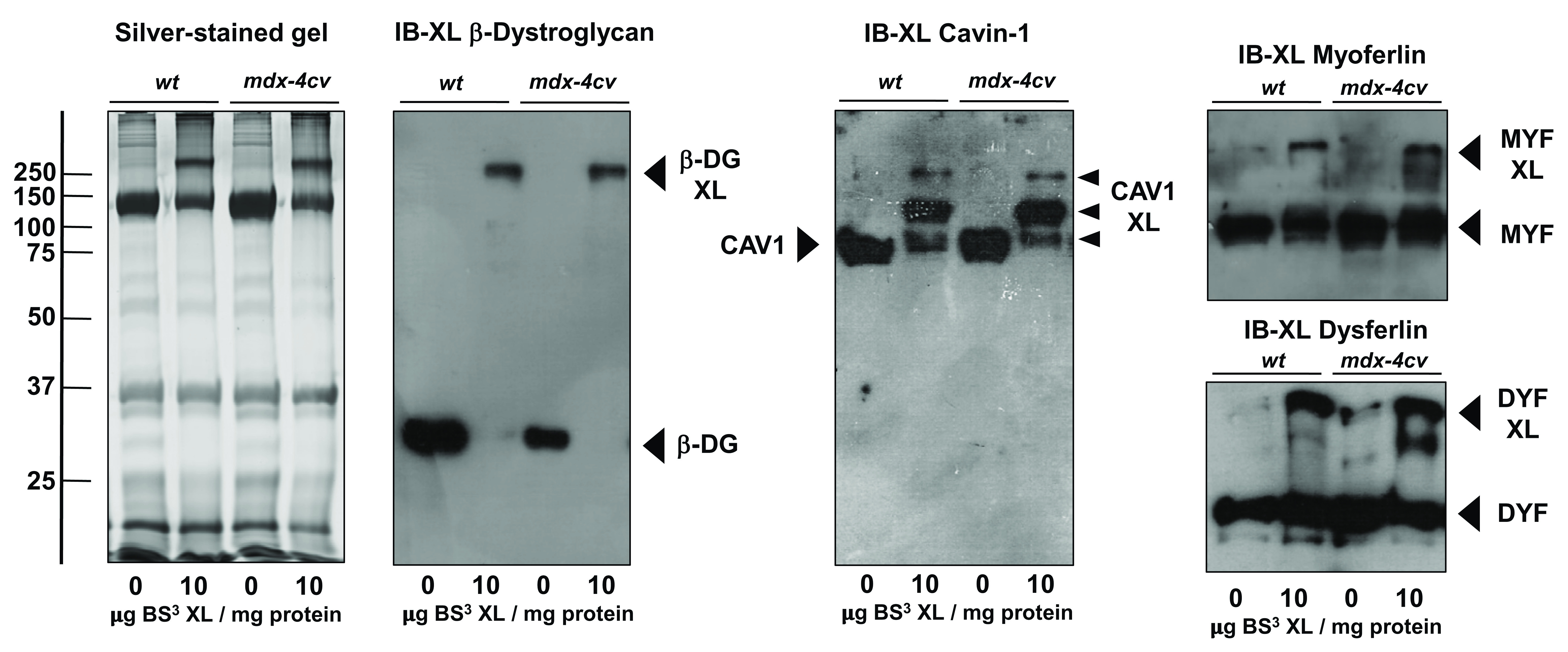
: In Duchenne muscular dystrophy, primary abnormalities in the membrane cytoskeletal protein dystrophin trigger the loss of sarcolemmal linkage between the extracellular matrix component laminin-211 and the intracellular cortical actin membrane cytoskeleton. The disintegration of the dystrophin-associated glycoprotein complex renders the plasma membrane of contractile fibres more susceptible to micro-rupturing, which is associated with abnormal calcium handling and impaired cellular signalling in dystrophinopathy. : The oligomerisation pattern of β-dystroglycan, an integral membrane protein belonging to the core dystrophin complex, was studied using immunoprecipitation and chemical crosslinking analysis. A homo-bifunctional and non-cleavable agent with water-soluble and amine-reactive properties was employed to study protein oligomerisation in normal versus dystrophin-deficient skeletal muscles. Crosslinker-induced protein oligomerisation was determined by a combination of gel-shift analysis and immunoblotting. : Although proteomics was successfully applied for the identification of dystroglycan as a key component of the dystrophin-associated glycoprotein complex in the muscle membrane fraction, mass spectrometric analysis did not efficiently recognize this relatively low-abundance protein after immunoprecipitation or chemical crosslinking. As an alternative approach, comparative immunoblotting was used to evaluate the effects of chemical crosslinking. Antibody decoration of the crosslinked microsomal protein fraction from wild type versus the mouse model of dystrophinopathy revealed oligomers that contain β-dystroglycan. The protein exhibited a comparable reduction in gel electrophoretic mobility in both normal and dystrophic samples. The membrane repair proteins dysferlin and myoferlin, which are essential components of fibre regeneration, as well as the caveolae-associated protein cavin-1, were also shown to exist in high-molecular mass complexes. : The muscular dystrophy-related reduction in the concentration of β-dystroglycan, which forms in conjunction with its extracellular binding partner α-dystroglycan a critical plasmalemmal receptor for laminin-211, does not appear to alter its oligomeric status. Thus, independent of direct interactions with dystrophin, this sarcolemmal glycoprotein appears to exist in a supramolecular assembly in muscle.
在杜兴氏肌营养不良症中,膜细胞骨架蛋白肌营养不良蛋白的原发性异常引发了细胞外基质成分层粘连蛋白-211与细胞内皮质肌动蛋白膜细胞骨架之间肌膜连接的丧失。肌营养不良蛋白相关糖蛋白复合物的解体使收缩纤维的质膜更容易发生微破裂,这与肌营养不良症中异常的钙处理和细胞信号传导受损有关。:使用免疫沉淀和化学交联分析研究了属于核心肌营养不良蛋白复合物的整合膜蛋白β-肌营养不良聚糖的寡聚化模式。一种具有水溶性和胺反应性的同型双功能且不可裂解的试剂被用于研究正常与缺乏肌营养不良蛋白的骨骼肌中的蛋白质寡聚化。交联剂诱导的蛋白质寡聚化通过凝胶迁移分析和免疫印迹相结合来确定。:尽管蛋白质组学已成功应用于鉴定肌营养不良聚糖是肌膜部分中肌营养不良蛋白相关糖蛋白复合物的关键成分,但质谱分析在免疫沉淀或化学交联后并不能有效地识别这种相对低丰度的蛋白质。作为一种替代方法,比较免疫印迹被用于评估化学交联的效果。来自野生型与肌营养不良症小鼠模型的交联微粒体蛋白部分的抗体标记揭示了含有β-肌营养不良聚糖的寡聚体。该蛋白在正常和营养不良样品中的凝胶电泳迁移率均有类似程度的降低。膜修复蛋白dysferlin和myoferlin是纤维再生的重要成分,以及小窝相关蛋白小窝蛋白-1,也被证明存在于高分子量复合物中。:与细胞外结合伴侣α-肌营养不良聚糖结合形成层粘连蛋白-211关键质膜受体的β-肌营养不良聚糖浓度的肌肉营养不良相关降低似乎并未改变其寡聚状态。因此,独立于与肌营养不良蛋白的直接相互作用,这种肌膜糖蛋白似乎以超分子组装形式存在于肌肉中。