Kou Bowen, Song Yang, Han Ye, Zhang Zepei, Miao Jun
Department of Spine Surgery, Tianjin Hospital of Tianjin University, Tianjin, China.
Department of Orthopaedics, Tianjin Medical University, Tianjin, China.
Ann Transl Med. 2022 Apr;10(7):415. doi: 10.21037/atm-22-969.
Orientation of the lumbar facet joints (FJs) in the transverse plane is associated with degenerative lumbar spine disease. However, there is a lack of measurements of the sagittal and coronal facet angles, and the effect of 3D facet angles on joint motion in the sitting position is unknown. The present study was to investigate the 3D orientation and motion characteristics of the FJ in the sitting position.
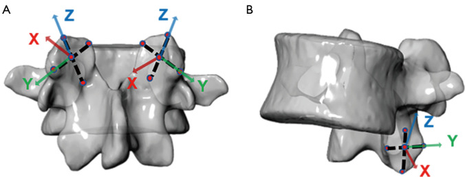
Dual fluoroscopic imaging system and computed tomography (CT) were used to determine the 3D orientation and kinematic characteristics of FJs. L3-S1 segments were studied in 10 asymptomatic participants (5 male and 5 female, age: 25-35 years, body mass index: 22.4±1.8). Angles of the facet in the sagittal, coronal, and axial planes, and the range of motion of the FJs in seated flexion and extension movements were measured.
The difference in sagittal facet angles between the 2 sides of the L3-S1 facet joints was not significant. The superior coronal facet angle on the left side of L5 was significantly smaller than that on the right side by 6.4° (P=0.01). The inferior transverse facet angle on the left side of L5 was greater than that on the right side by 7.1; the results were not statistically significantly different. In the sitting position, the range of motion of the left and right sides of L5-S1 differed significantly, with the right side being 5.5° (P=0.004) and 11.7° (P=0.026) greater than the left side in the sagittal and coronal planes, respectively. There was a correlation between mobility and the 3D orientation angle of the FJs in each segment.
Quantification of the 3D orientation of the lumbar spine FJs provides new perspectives to study the kinematics of the lumbar spine and the etiology of lumbar degenerative diseases. In sitting flexion and extension movements, there is a significant difference in the left-right lateral mobility of the FJs of the L5-S1 segments. With the exception of the transverse facet angle of the lumbar spine FJs, the sagittal and coronal facet angles also have an effect on lumbar spine mobility.
腰椎小关节(FJ)在横平面的方向与腰椎退行性疾病相关。然而,矢状面和冠状面小关节角度的测量尚缺乏,并且三维小关节角度对坐位时关节运动的影响未知。本研究旨在探讨坐位时FJ的三维方向和运动特征。
使用双荧光透视成像系统和计算机断层扫描(CT)来确定FJ的三维方向和运动学特征。对10名无症状参与者(5名男性和5名女性,年龄:25 - 35岁,体重指数:22.4±1.8)的L3 - S1节段进行研究。测量矢状面、冠状面和轴平面的小关节角度,以及坐位屈伸运动中FJ的运动范围。
L3 - S1小关节两侧矢状面小关节角度差异不显著。L5左侧上冠状面小关节角度比右侧显著小6.4°(P = 0.01)。L5左侧下横切面小关节角度比右侧大7.1°;结果无统计学显著差异。在坐位时,L5 - S1左右两侧的运动范围有显著差异,矢状面和冠状面右侧分别比左侧大5.5°(P = 0.004)和11.7°(P = 0.026)。每个节段FJ的活动度与三维方向角之间存在相关性。
腰椎FJ三维方向的量化为研究腰椎运动学和腰椎退行性疾病的病因提供了新的视角。在坐位屈伸运动中,L5 - S1节段FJ的左右侧向活动度存在显著差异。除腰椎FJ的横切面角度外,矢状面和冠状面小关节角度也对腰椎活动度有影响。