Pathology Division, United States Army Medical Research Institute of Infectious Diseases, Frederick, Maryland, United States of America.
Virology Division, United States Army Medical Research Institute of Infectious Diseases, Frederick, Maryland, United States of America.
PLoS Negl Trop Dis. 2022 May 9;16(5):e0010081. doi: 10.1371/journal.pntd.0010081. eCollection 2022 May.
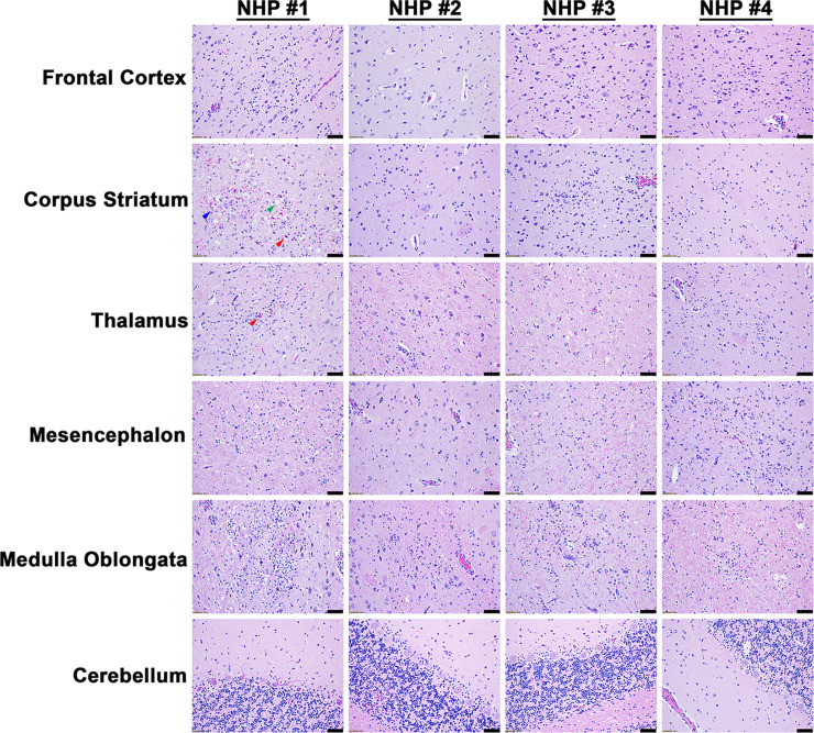
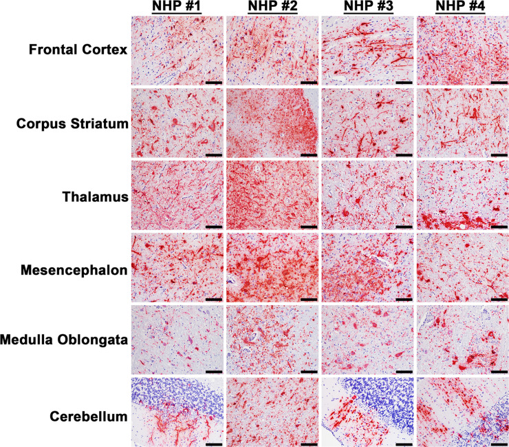
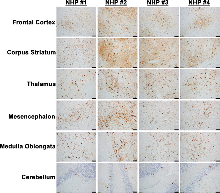
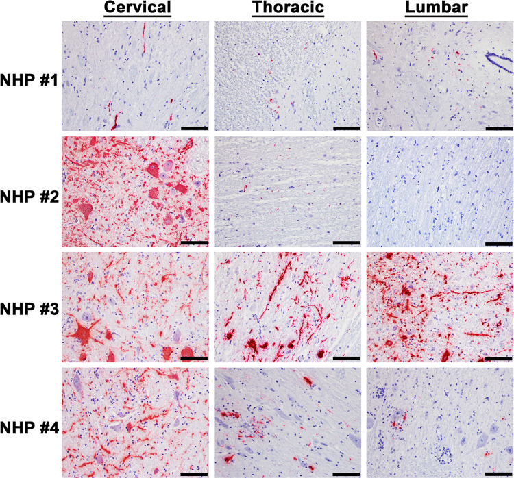
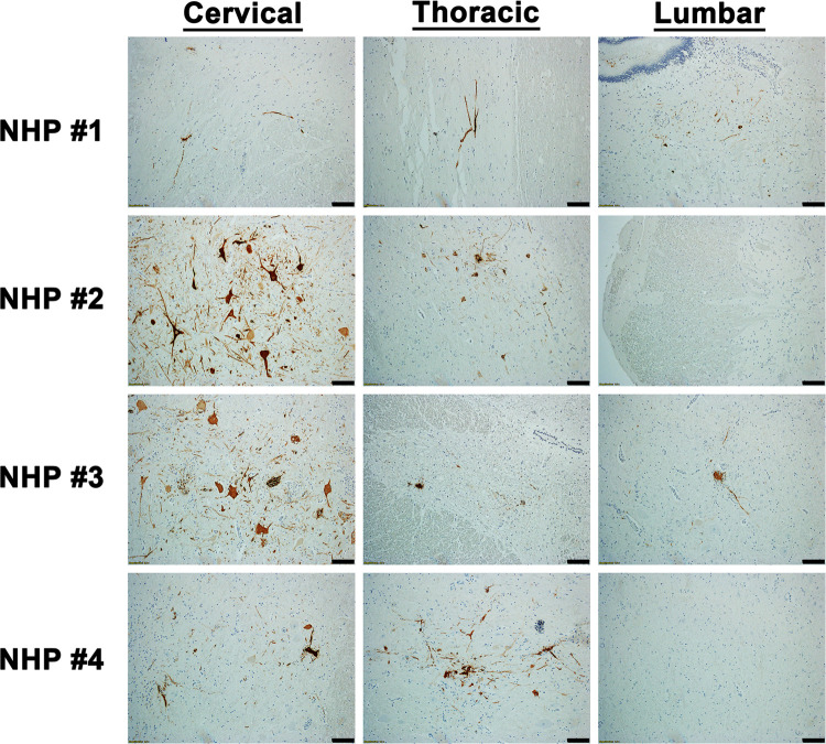
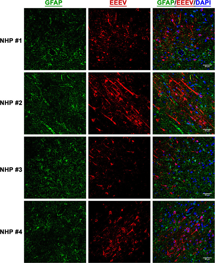
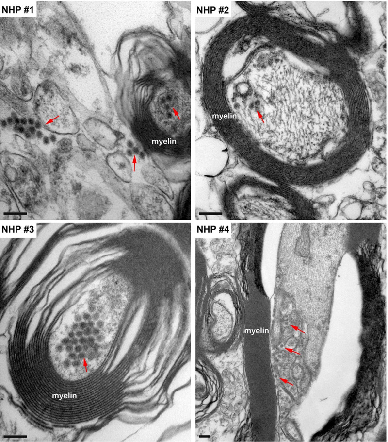
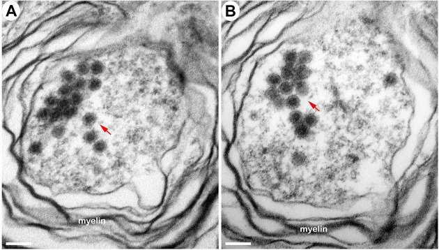
Eastern equine encephalitis virus (EEEV) is mosquito-borne virus that produces fatal encephalitis in humans. We recently conducted a first of its kind study to investigate EEEV clinical disease course following aerosol challenge in a cynomolgus macaque model utilizing the state-of-the-art telemetry to measure critical physiological parameters. Here, we report the results of a comprehensive pathology study of NHP tissues collected at euthanasia to gain insights into EEEV pathogenesis. Viral RNA and proteins as well as microscopic lesions were absent in the visceral organs. In contrast, viral RNA and proteins were readily detected throughout the brain including autonomic nervous system (ANS) control centers and spinal cord. However, despite presence of viral RNA and proteins, majority of the brain and spinal cord tissues exhibited minimal or no microscopic lesions. The virus tropism was restricted primarily to neurons, and virus particles (~61-68 nm) were present within axons of neurons and throughout the extracellular spaces. However, active virus replication was absent or minimal in majority of the brain and was limited to regions proximal to the olfactory tract. These data suggest that EEEV initially replicates in/near the olfactory bulb following aerosol challenge and is rapidly transported to distal regions of the brain by exploiting the neuronal axonal transport system to facilitate neuron-to-neuron spread. Once within the brain, the virus gains access to the ANS control centers likely leading to disruption and/or dysregulation of critical physiological parameters to produce severe disease. Moreover, the absence of microscopic lesions strongly suggests that the underlying mechanism of EEEV pathogenesis is due to neuronal dysfunction rather than neuronal death. This study is the first comprehensive investigation into EEEV pathology in a NHP model and will provide significant insights into the evaluation of countermeasure.
东部马脑炎病毒(EEEV)是一种通过蚊子传播的病毒,可导致人类致命脑炎。我们最近进行了首例此类研究,利用最先进的遥测技术测量关键生理参数,调查食蟹猴模型中气溶胶挑战后 EEEV 的临床疾病过程。在这里,我们报告了对安乐死时收集的 NHPT 组织进行的全面病理学研究的结果,以深入了解 EEEV 的发病机制。内脏器官中未检测到病毒 RNA 和蛋白质以及微观病变。相比之下,在大脑中,包括自主神经系统(ANS)控制中心和脊髓在内,都可以轻易检测到病毒 RNA 和蛋白质。然而,尽管存在病毒 RNA 和蛋白质,但大多数大脑和脊髓组织的微观病变很少或没有。病毒的嗜性主要局限于神经元,病毒颗粒(~61-68nm)存在于神经元的轴突内和整个细胞外空间内。然而,大多数大脑中的病毒复制是不存在或很少的,仅限于靠近嗅束的区域。这些数据表明,EEEV 在气溶胶挑战后最初在嗅球内/附近复制,并通过利用神经元轴突运输系统迅速运输到大脑的远端区域,从而促进神经元之间的传播。一旦进入大脑,病毒就可以进入 ANS 控制中心,可能导致关键生理参数的中断和/或失调,从而产生严重疾病。此外,微观病变的缺失强烈表明,EEEV 发病机制的潜在机制是神经元功能障碍而不是神经元死亡。这项研究是对食蟹猴模型中 EEEV 病理学的首次全面调查,将为评估对策提供重要见解。