Mahendiran Dharmasivam, Amuthakala Sethu, Bhuvanesh Nattamai S P, Kumar Raju Senthil, Rahiman Aziz Kalilur
Post-Graduate and Research Department of Chemistry, The New College (Autonomous) Chennai 600 014 India
Department of Chemistry, Texas A & M University College Station TX 77842 USA.
RSC Adv. 2018 May 9;8(30):16973-16990. doi: 10.1039/c8ra00954f. eCollection 2018 May 3.
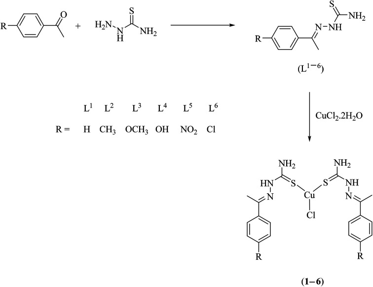
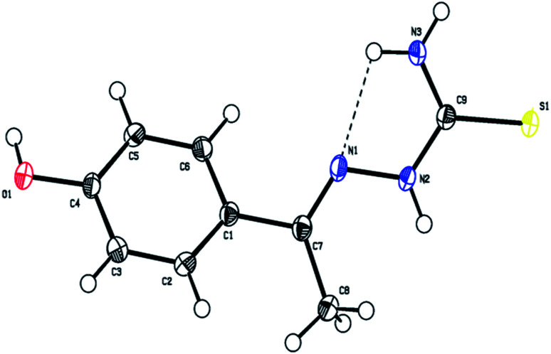
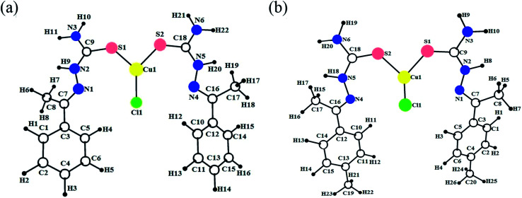
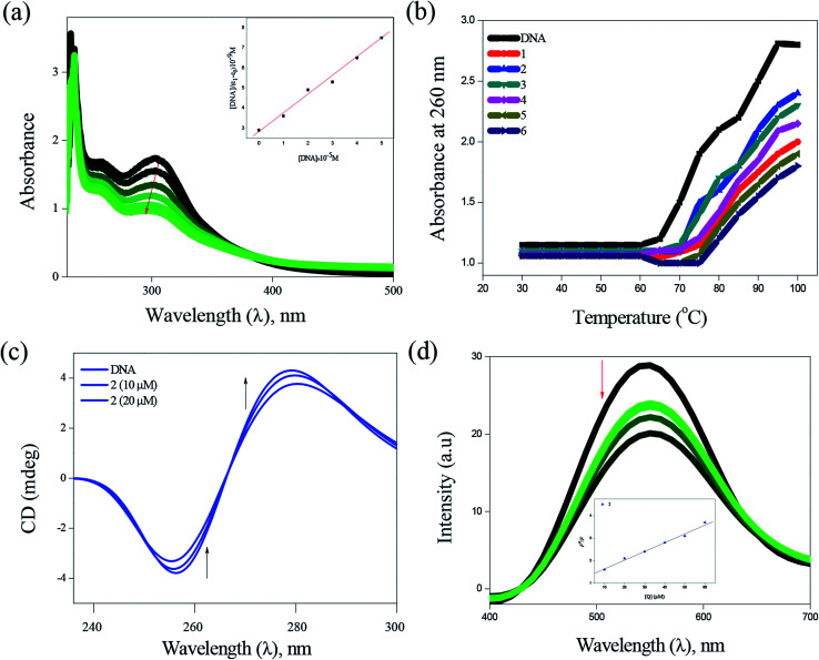
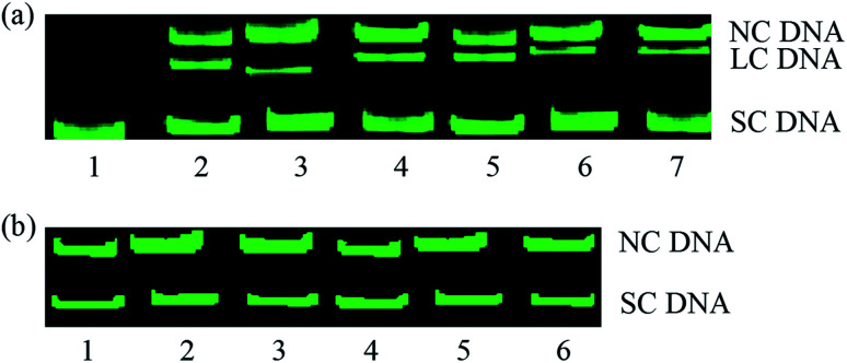
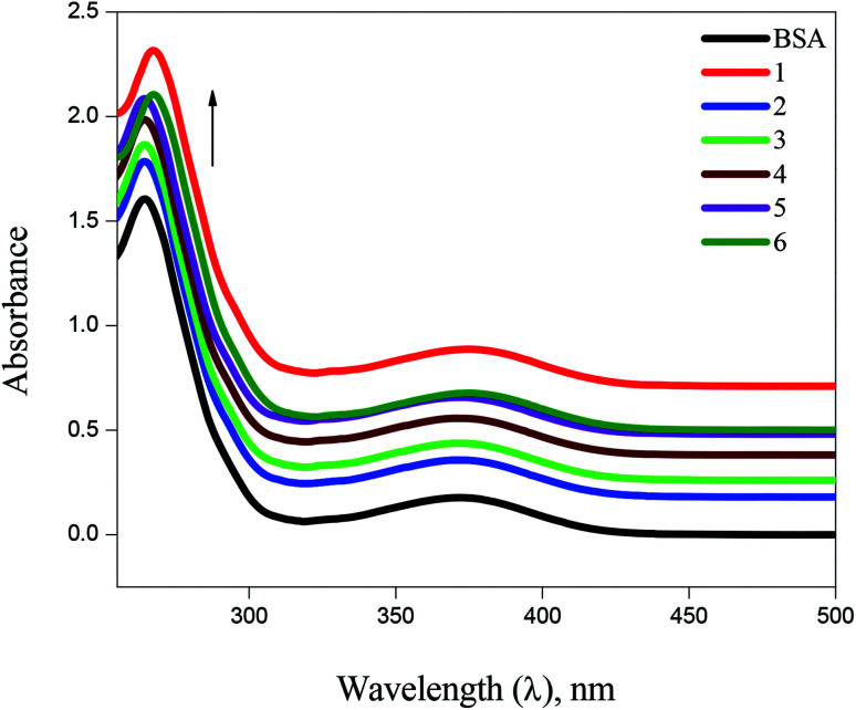
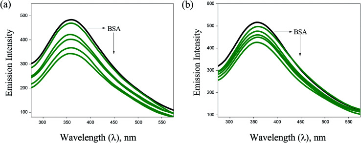
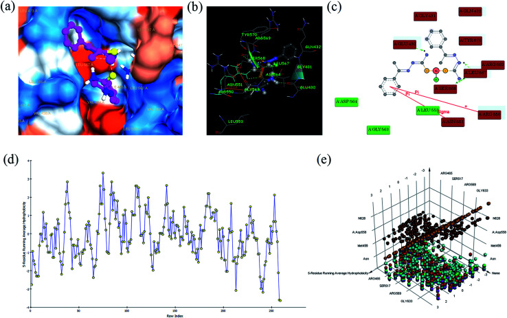
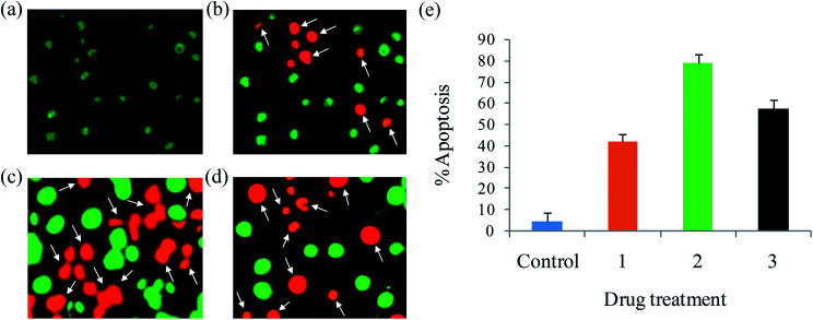
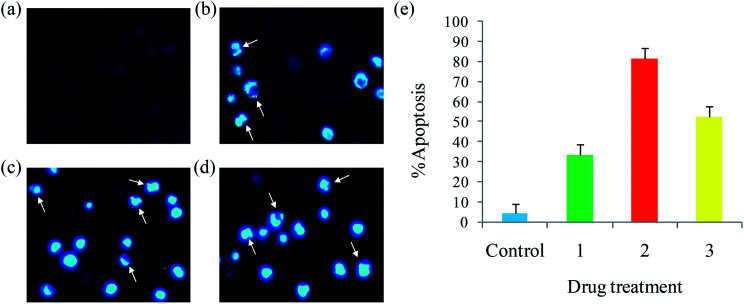
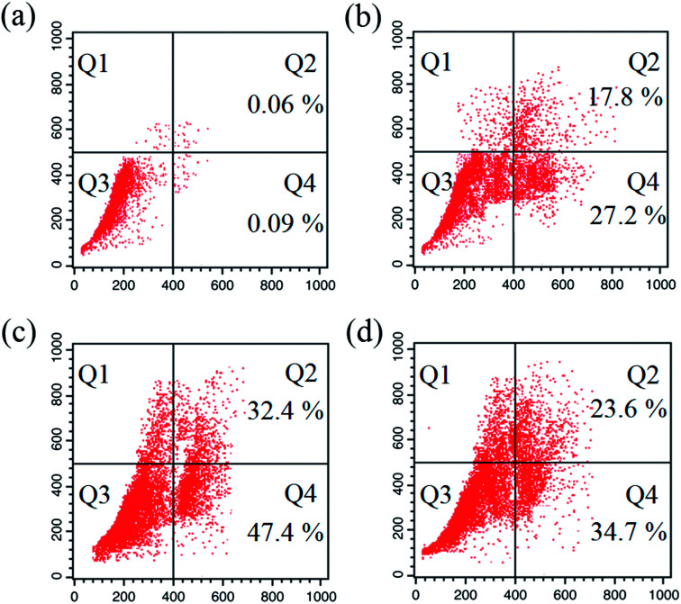
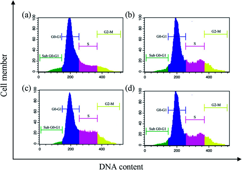
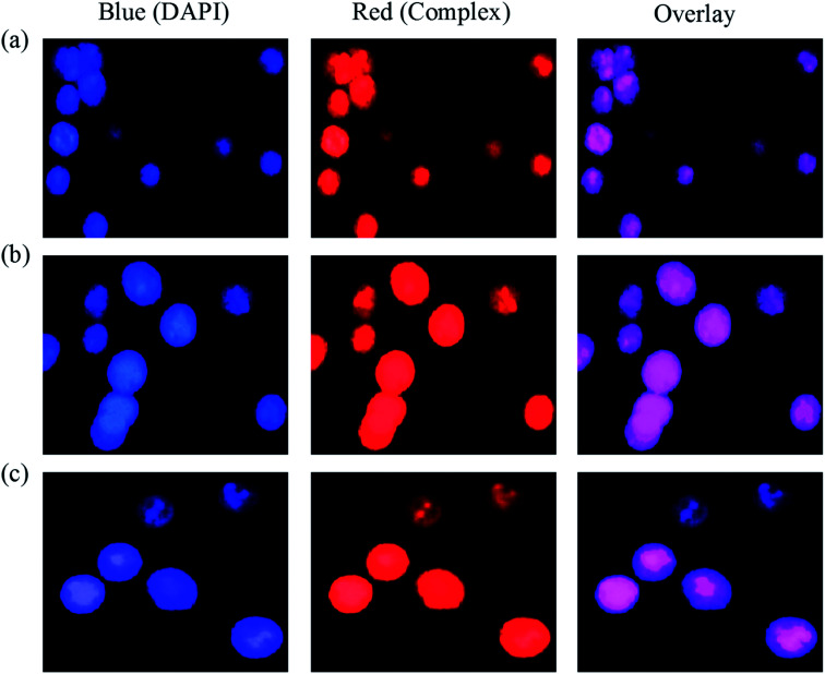
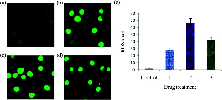
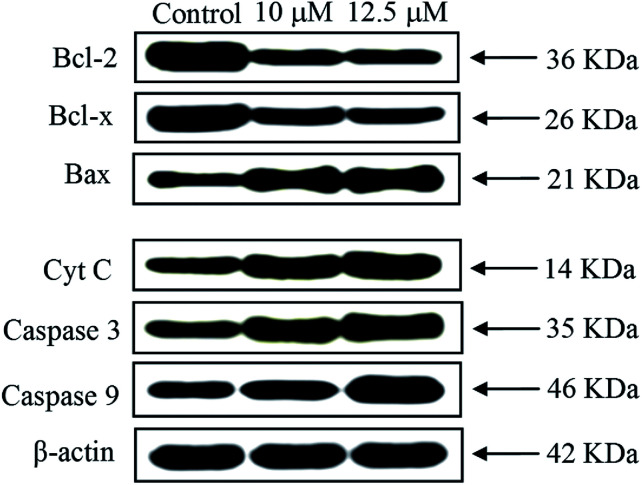
A series of six new bis(thiosemicarbazone)copper(i) complexes of the type [Cu(L)Cl] (1-6) have been synthesized and characterized. The molecular structure of the ligand L was determined by the single crystal XRD method. All the complexes adopted trigonal planar (Y-shaped) geometry. All the complexes strongly bind with CT-DNA intercalative mode, which was further supported by molecular docking studies. Further, the complexes were effectively bind with BSA as observed by UV-Vis and fluorescence spectra. All the complexes effectively cleave pBR322 DNA through hydrolytic pathway as evidenced from T4 ligase experiments. All the complexes interact with the anticancer receptor focal adhesion kinase (FAK) electrostatic, van der Waals, hydrogen bonding, σ-π and π-π interactions. cytotoxicity of the complexes were assessed by MTT assay against four cancer cell lines such as human breast adenocarcinoma (MCF-7), cervical (HeLa), epithelioma (Hep-2) and Ehrlich ascites carcinoma (EAC), and two normal cell lines namely normal human dermal fibroblasts (NHDF) and L6 myotubes with respect to the commercially used anticancer drug cisplatin. All the complexes induce apoptosis in EAC cells, which was confirmed by AO/EB, Hoechst 33258 and PI staining methods. The complexes block cell cycle progression of EAC cells in S phase (DNA synthesis). The cellular uptake studies confirmed the ability of the complexes to go into the cytoplasm and accumulation in the cell nuclei. In the anticancer studies, the complexes significantly reduce the tumour volume in female Swiss albino mice. Overall, our results ensure the role of thiosemicarbazone-based copper(i) complexes as prospective anticancer agents, induction of apoptosis and S phase arrest with the mitochondrial controlled pathway.
已合成并表征了一系列六种新型的[Cu(L)Cl]型双(硫代半卡巴腙)铜(I)配合物(1-6)。通过单晶XRD方法确定了配体L的分子结构。所有配合物均采用三角平面(Y形)几何结构。所有配合物均以嵌入模式与CT-DNA强烈结合,分子对接研究进一步证实了这一点。此外,通过紫外可见光谱和荧光光谱观察到,这些配合物能有效地与牛血清白蛋白结合。T4连接酶实验证明,所有配合物均通过水解途径有效切割pBR322 DNA。所有配合物均通过静电、范德华力、氢键、σ-π和π-π相互作用与抗癌受体粘着斑激酶(FAK)相互作用。通过MTT法评估了这些配合物对四种癌细胞系(如人乳腺腺癌(MCF-7)、宫颈癌(HeLa)、上皮瘤(Hep-2)和艾氏腹水癌(EAC))以及两种正常细胞系(正常人皮肤成纤维细胞(NHDF)和L6肌管)相对于市售抗癌药物顺铂的细胞毒性。所有配合物均能诱导EAC细胞凋亡,AO/EB、Hoechst 33258和PI染色方法证实了这一点。这些配合物可阻断EAC细胞在S期(DNA合成)的细胞周期进程。细胞摄取研究证实了这些配合物进入细胞质并在细胞核中积累的能力。在抗癌研究中,这些配合物显著减小了雌性瑞士白化小鼠的肿瘤体积。总体而言,我们的结果证实了基于硫代半卡巴腙的铜(I)配合物作为潜在抗癌药物的作用,通过线粒体控制途径诱导凋亡和S期阻滞。