Song Weiling, Zhang Nan, Luan Zhenzhu, Zhang Xiaoru, He Peng
Key Laboratory of Sensor Analysis of Tumor Marker, Ministry of Education, College of Chemistry and Molecular Engineering, Qingdao University of Science and Technology Qingdao 266042 P. R. China
RSC Adv. 2018 Apr 23;8(27):15248-15252. doi: 10.1039/c8ra01799a. eCollection 2018 Apr 18.
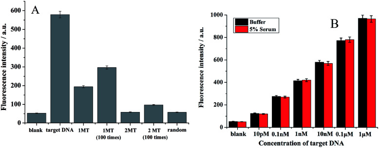
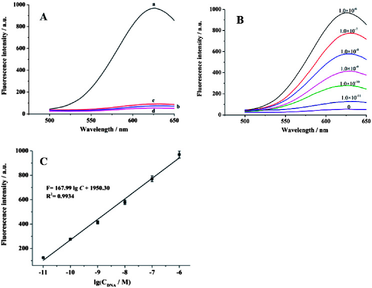
A novel detection method based on the cation-exchange reaction of CuS nanoparticles (CuS NPs) combined with poly T-templated fluorescent Cu nanoparticles (Cu NPs) was developed. First, CuS NPs-magnetic bead conjugates were prepared through the hybridization of DNA. Competition with target DNA resulted in the release of CuS NPs, and exonuclease III catalysis could lead to recycling of the target DNA. Then, the CuS NPs released into the supernatant were subjected to a cation-exchange reaction after the addition of AgNO. The obtained Cu could form fluorescent Cu NPs using poly T DNA as a template. The fluorescence intensity of the Cu NPs could be used to determine the concentration of the target DNA. To further increase the detection sensitivity, two types of DNA decorated magnetic beads were used. After Exo III digestion for two cycle processes, more CuS NPs entered the supernatant. Hence, a stronger fluorescence intensity was found after the cation-exchange reaction and the formation of fluorescent Cu NPs. The developed method is convenient and low cost with good sensitivity and selectivity.
开发了一种基于硫化铜纳米颗粒(CuS NPs)与聚T模板荧光铜纳米颗粒(Cu NPs)阳离子交换反应的新型检测方法。首先,通过DNA杂交制备CuS NPs-磁珠缀合物。与靶DNA竞争导致CuS NPs释放,外切核酸酶III催化可导致靶DNA循环利用。然后,加入AgNO后,释放到上清液中的CuS NPs进行阳离子交换反应。得到的Cu可以使用聚T DNA作为模板形成荧光Cu NPs。Cu NPs的荧光强度可用于测定靶DNA的浓度。为了进一步提高检测灵敏度,使用了两种类型的DNA修饰磁珠。经过两个循环过程的Exo III消化后,更多的CuS NPs进入上清液。因此,在阳离子交换反应和荧光Cu NPs形成后发现荧光强度更强。所开发的方法方便、低成本,具有良好的灵敏度和选择性。