Department of Gastrointestinal Surgery & Department of Gastric and Colorectal Surgical Oncology, Zhongnan Hospital of Wuhan University, Wuhan, Hubei, 430071, China.
Department of obstetrics and gynecology, Guangzhou Women and Children's Medical Center, Guangzhou, Guangdong, 510623, China.
Int J Biol Sci. 2022 Apr 24;18(7):3082-3101. doi: 10.7150/ijbs.70524. eCollection 2022.
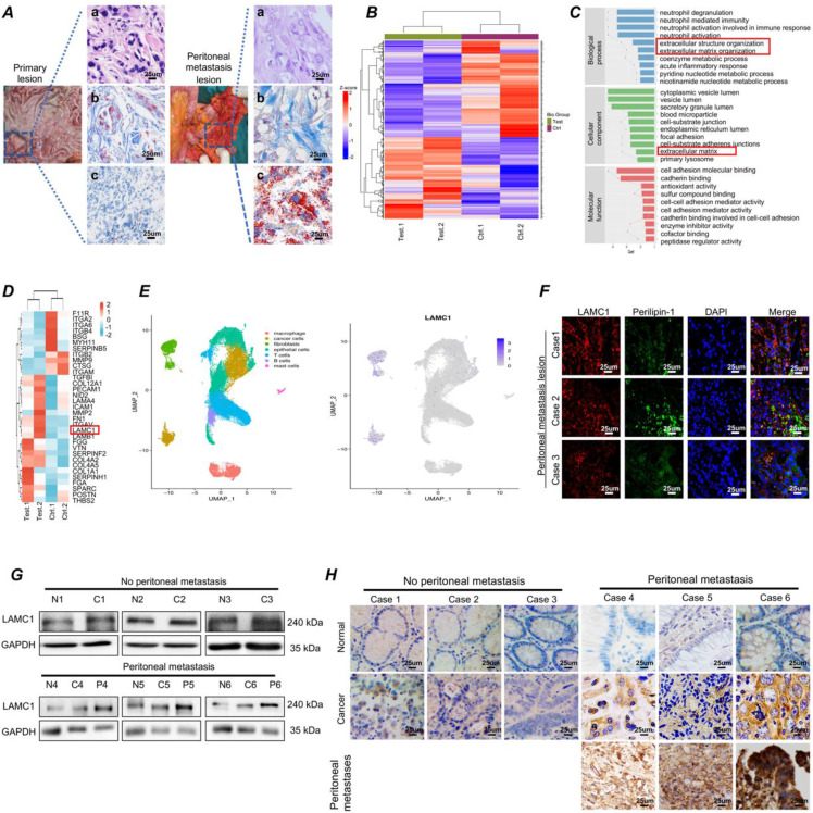
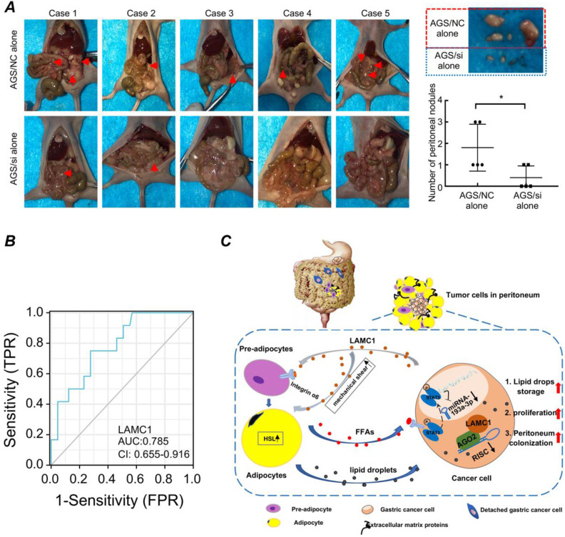
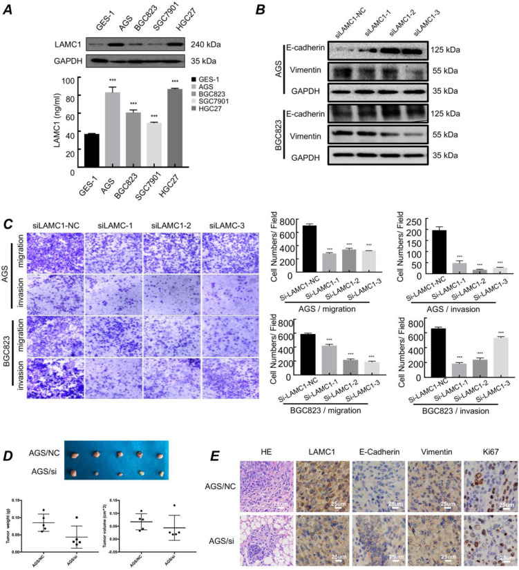
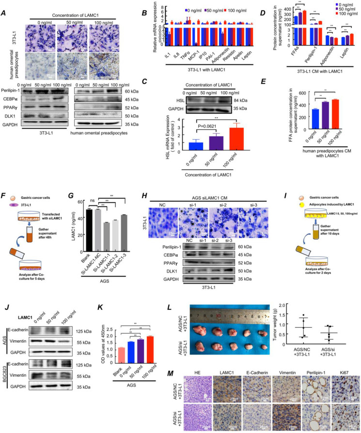
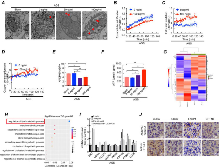
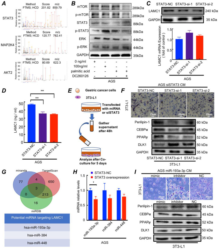
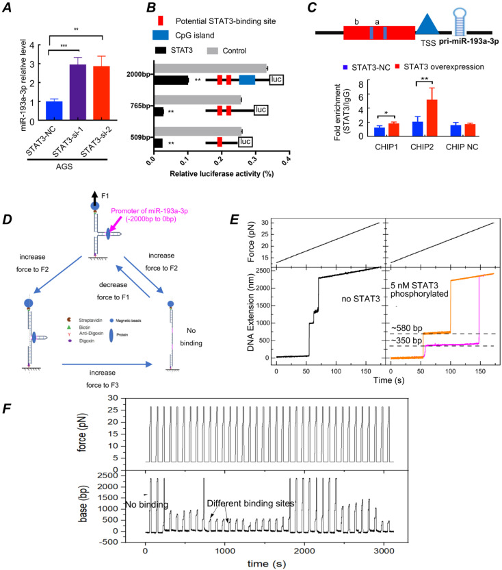
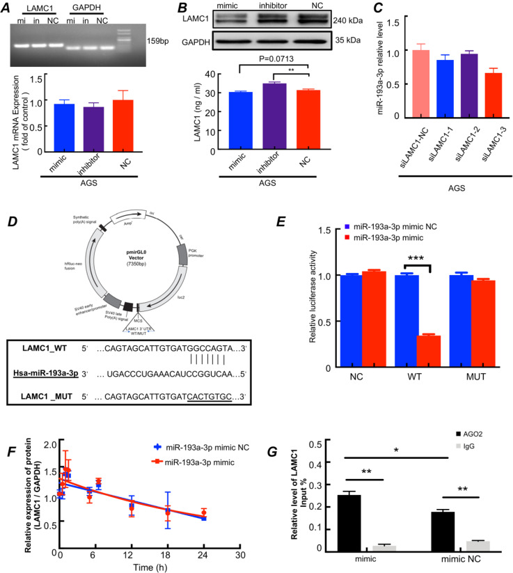
Gastric cancer is anatomically proximal to peritoneum. Gastric cancer peritoneal metastasis is a complex biological process which is corresponded with disharmony within dysfunctional adipose tissue and metabolism reprogramming. Laminin gamma 1 (LAMC1) is highly expressed in cancer cells of peritoneal metastatic sites, however, the mechanism of LAMC1-metiated gastric cancer metastases to adipose tissue-rich peritoneum remains unclear. In our study, immunohistochemical staining, single cell sequencing, a co-culture model, luciferase reporter, RNA immunoprecipitation (RIP), Chromatin immunoprecipitation (CHIP) and single-molecular magnetic tweezers assays were conducted, and our results showed that LAMC1 related to Perilipin-1 content was highly expressed in peritoneal metastatic sites and mainly secreted by tumor cells. Gastric cancer cells secreted LAMC1 in an autocrine manner to detached from the primary site and promoted preadipocytes mature, rupture and release of free fatty acids (FFAs) in the peritoneal microenvironment to form pre-metastatic niche by the paracrine pathway. Reversely, differentiated preadipocyte-derived conditioned medium inhibited glycolysis and enhanced fatty acid oxidation (FAO) rate to promote cell proliferation, mesenchymal-epithelial transformation which led to tumor peritoneal colonization. In terms of biological mechanisms, one of differentiated preadipocyte-derived FFAs, palmitic acid-activated STAT3 inhibited miR-193a-3p by binding to its promoter directly; Using single-molecular magnetic tweezers, this binding manner was proved to be stable, reversable and ATP-dependent. Moreover, miR-193a-3p regulated LAMC1 in a post-translational manner. Furthermore, high LAMC1 expression in serum predicted a higher risk of peritoneal metastasis. In conclusion, our results illustrated that palmitic acid/p-STAT3/miR-193a-3p/LAMC1 pathway promotes preadipocyte differentiation, pre-metastatic niche formation and gastric cancer cell colonization to peritoneum.
胃癌在解剖学上与腹膜相邻。胃癌腹膜转移是一个复杂的生物学过程,与功能失调的脂肪组织和代谢重编程不协调有关。层粘连蛋白 γ1(LAMC1)在腹膜转移部位的癌细胞中高表达,然而,LAMC1 介导的胃癌转移到富含脂肪的腹膜的机制尚不清楚。在我们的研究中,进行了免疫组织化学染色、单细胞测序、共培养模型、荧光素酶报告基因、RNA 免疫沉淀(RIP)、染色质免疫沉淀(CHIP)和单分子磁镊实验,结果表明 LAMC1 与 perilipin-1 含量在腹膜转移部位高表达,主要由肿瘤细胞分泌。胃癌细胞以自分泌方式分泌 LAMC1,从而从原发部位脱离,并促进前脂肪细胞成熟,在腹膜微环境中破裂并释放游离脂肪酸(FFAs),通过旁分泌途径形成前转移龛。相反,分化的前脂肪细胞衍生的条件培养基抑制糖酵解,增强脂肪酸氧化(FAO)率,促进细胞增殖、上皮-间充质转化,导致肿瘤腹膜定植。就生物学机制而言,分化的前脂肪细胞衍生的 FFAs 之一,棕榈酸激活的 STAT3 通过直接结合其启动子抑制 miR-193a-3p;使用单分子磁镊,证明这种结合方式是稳定的、可逆的和 ATP 依赖性的。此外,miR-193a-3p 以翻译后方式调节 LAMC1。此外,血清中高 LAMC1 表达预示着腹膜转移的风险更高。总之,我们的结果表明,棕榈酸/p-STAT3/miR-193a-3p/LAMC1 通路促进前脂肪细胞分化、前转移龛形成和胃癌细胞向腹膜的定植。