Sha Yang, Yang Weimin, Li Sanyang, Yao Liangbo, Li Haoyi, Cheng Lisheng, Yan Hua, Cao Weiyu, Tan Jing
State Key Laboratory of Organic-Inorganic Composites, College of Material Science and Engineering, Beijing University of Chemical Technology Beijing 100029 China
The Key Laboratory of Education Ministry on Carbon Fibre and Functional Polymer, Beijing University of Chemical Technology Beijing 100029 China.
RSC Adv. 2018 Mar 23;8(21):11543-11550. doi: 10.1039/c8ra00497h. eCollection 2018 Mar 21.
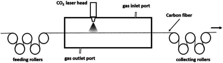
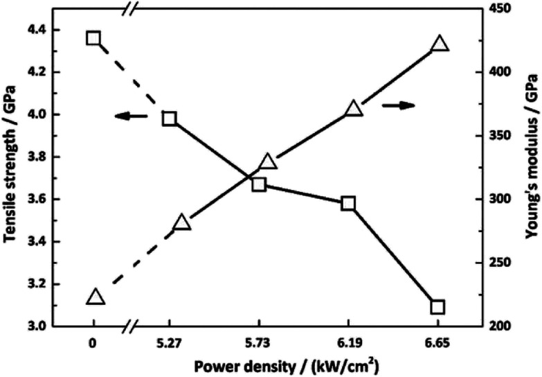
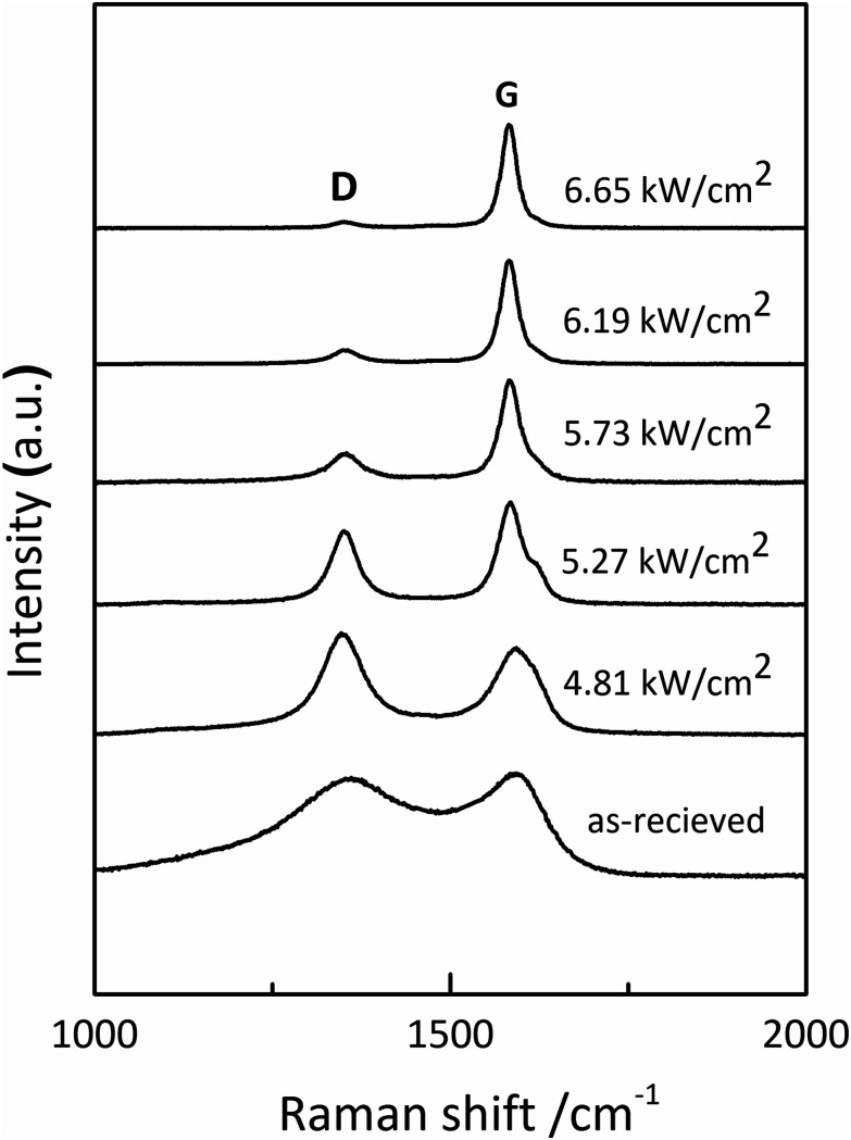
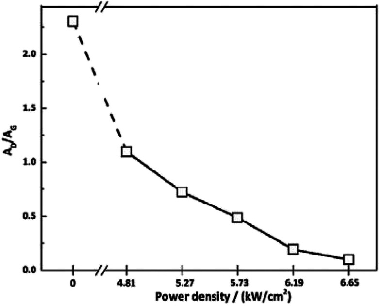
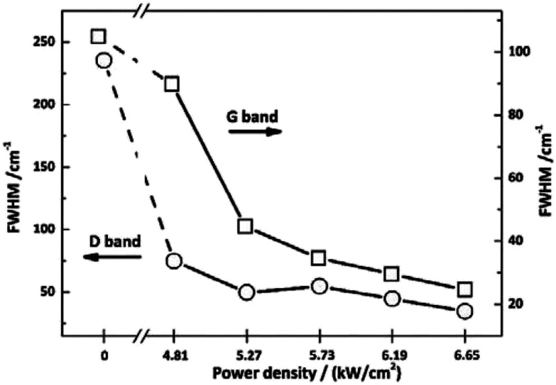
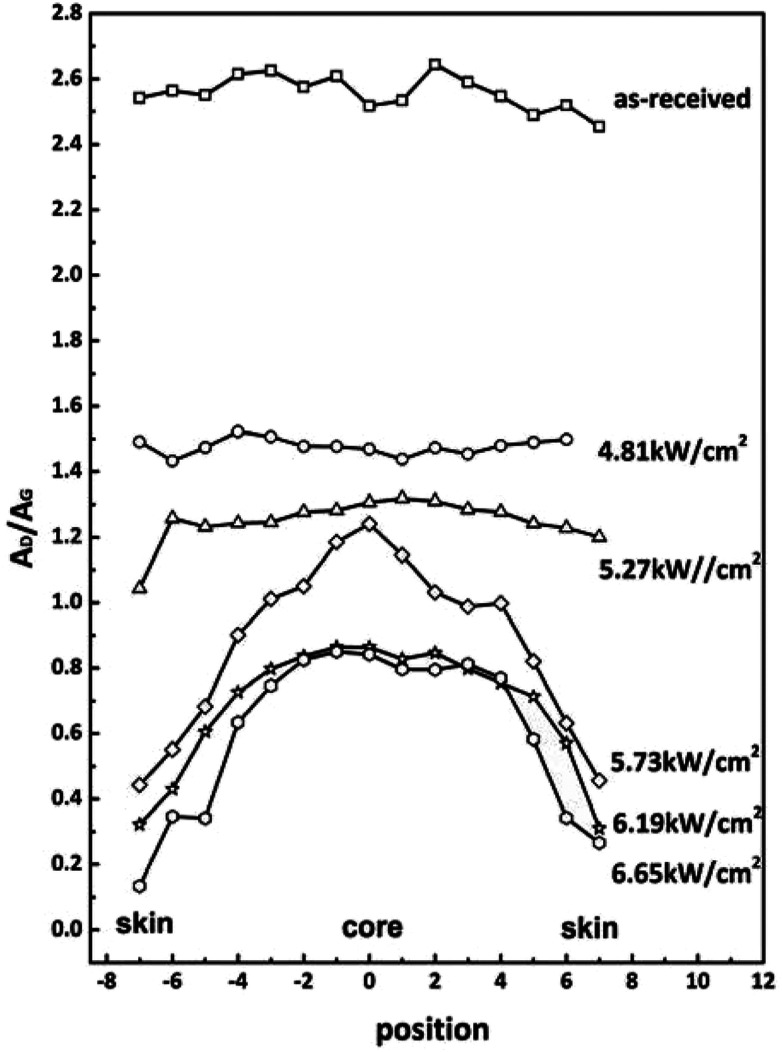
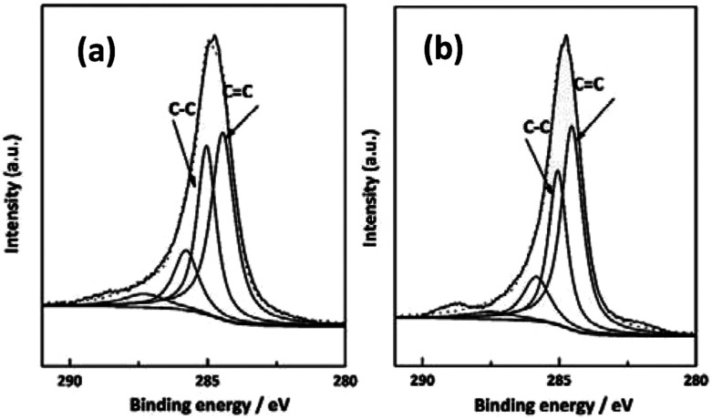
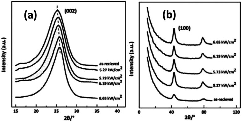
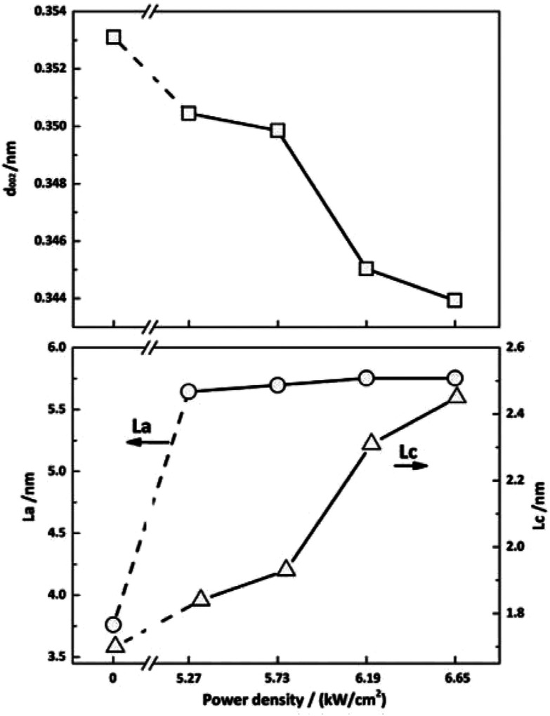
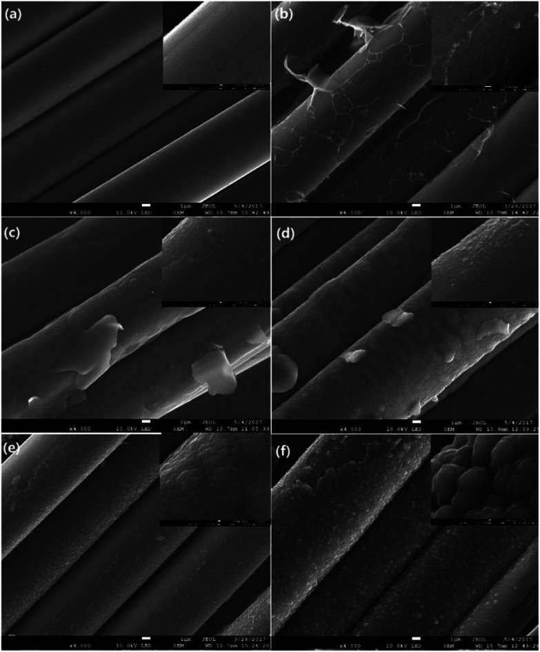
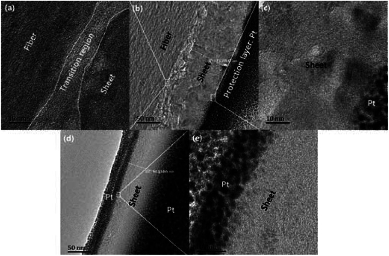
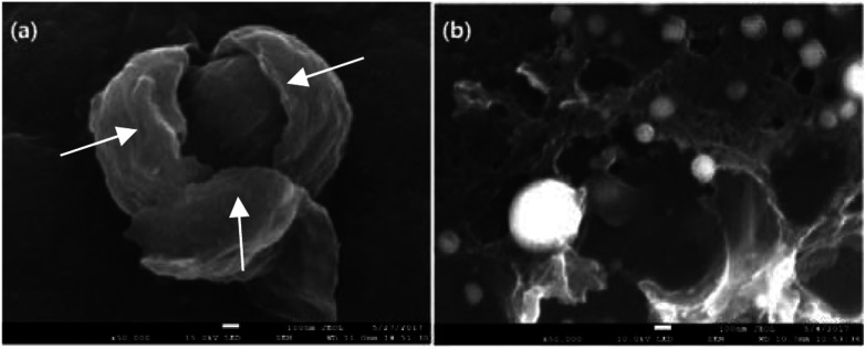
Laser induced graphitization of polyacrylonitrile-based carbon fibers (CFs) was carried out in a self-designed furnace with a CO laser source. The microstructures combined with mechanical properties of the irradiated CFs were measured by Raman spectroscopy, X-ray photoelectron spectroscopy (XPS), wide-angle X-ray diffraction (WAXD), scanning electron microscopy (SEM), high resolution transmission electron microscopy (HRTEM) and single filament tensile test, respectively. The results exhibited that the hierarchical structures of CFs showed different responses to the CO laser. After laser graphitization, the surface and cross-section structure were characterized by Raman spectroscopy. As the power density increased, a profound increase of graphitization degree happened and obvious skin-core structures were observed. Furthermore, the results of XPS measurements indicated that the irradiated CFs showed more conjugated structures. For crystallite structure, the interlayer spacing of the (002) lattice decreased and the thickness of crystallite increased after graphitization. The size of the (002) lattice parallel to the fiber axis changed slightly. The surface morphology was also investigated by SEM, sheet structures and particles could be observed on the surface of CFs. This was attributed to fast energy addition of laser and the characteristics of the material. Further HRTEM investigation revealed that the sheet structure is multilayered graphene. The Young's modulus of irradiated fibers showed obvious improvements compared to that of as-received ones.
采用自行设计的带有CO激光源的炉体,对聚丙烯腈基碳纤维(CFs)进行激光诱导石墨化处理。分别通过拉曼光谱、X射线光电子能谱(XPS)、广角X射线衍射(WAXD)、扫描电子显微镜(SEM)、高分辨率透射电子显微镜(HRTEM)和单丝拉伸试验,对辐照后CFs的微观结构和力学性能进行了测量。结果表明,CFs的分级结构对CO激光表现出不同的响应。激光石墨化后,通过拉曼光谱对其表面和横截面结构进行了表征。随着功率密度的增加,石墨化程度显著提高,且观察到明显的皮芯结构。此外,XPS测量结果表明,辐照后的CFs表现出更多的共轭结构。对于微晶结构,石墨化后(002)晶格的层间距减小,微晶厚度增加。平行于纤维轴的(002)晶格尺寸变化不大。还通过SEM研究了表面形貌,在CFs表面可观察到片状结构和颗粒。这归因于激光的快速能量输入和材料的特性。进一步的HRTEM研究表明,片状结构为多层石墨烯。与未处理的纤维相比,辐照后纤维的杨氏模量有明显提高。