• 文献检索
  • 文档翻译
  • 深度研究
  • 学术资讯
  • Suppr Zotero 插件Zotero 插件
  • 邀请有礼
  • 套餐&价格
  • 历史记录
应用&插件
Suppr Zotero 插件Zotero 插件浏览器插件Mac 客户端Windows 客户端微信小程序
定价
高级版会员购买积分包购买API积分包
服务
文献检索文档翻译深度研究API 文档MCP 服务
关于我们
关于 Suppr公司介绍联系我们用户协议隐私条款
关注我们

Suppr 超能文献

核心技术专利:CN118964589B侵权必究
粤ICP备2023148730 号-1Suppr @ 2026

文献检索

告别复杂PubMed语法,用中文像聊天一样搜索,搜遍4000万医学文献。AI智能推荐,让科研检索更轻松。

立即免费搜索

文件翻译

保留排版,准确专业,支持PDF/Word/PPT等文件格式,支持 12+语言互译。

免费翻译文档

深度研究

AI帮你快速写综述,25分钟生成高质量综述,智能提取关键信息,辅助科研写作。

立即免费体验

通过非侵入性评估确定门静脉高压症的严重程度:肝脏实质的声学结构定量分析。

The severity of portal hypertension by a non-invasive assessment: acoustic structure quantification analysis of liver parenchyma.

机构信息

Department of Ultrasound Diagnosis, Tangdu Hospital, The Fourth Military Medical University, Xin Si Road, Ba Qiao District, Xi'an, China.

Department of Ultrasound Diagnostics, General Hospital of Tibet Military Region, Lhasa, China.

出版信息

BMC Med Imaging. 2022 May 12;22(1):85. doi: 10.1186/s12880-022-00817-2.

DOI:10.1186/s12880-022-00817-2
PMID:35550032
原文链接:https://pmc.ncbi.nlm.nih.gov/articles/PMC9097305/
Abstract

BACKGROUND

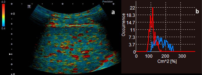
Acoustic structure quantification (ASQ) has been applied to evaluate liver histologic changes by analyzing the speckle pattern seen on B-mode ultrasound. We aimed to assess the severity of portal hypertension (PHT) through hepatic ultrasonography.

METHODS

Sixty patients diagnosed with PHT and underwent surgical treatment with portosystemic shunts were enrolled. Portal pressure (PP) was measured intraoperatively. Patients were divided into subgroups according to the severity of gastroesophageal varices and Child-Pugh class. Three difference ratio (C) values on ASQ histogram mode were analyzed for their relationships with PP, degree of gastroesophageal varices and Child-Pugh liver function. Thirty healthy volunteers matched with the patients for gender and age were enrolled as controls. Comparisons among groups and correlation of the parameters with PP were analyzed. Area under the receive operating characteristic curve was used to evaluate the predicting value of ASQ parameters.

RESULTS

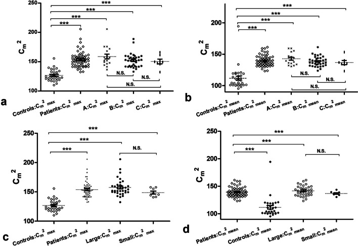
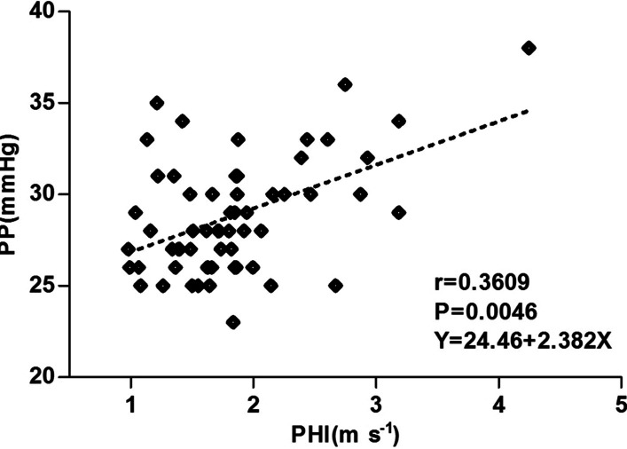
In the patients, the ASQ parameters peak C (C), mean C (C) and the highest occurred C value of the obtained red curve (RC) were all greatly increased (P < 0.0001, P < 0.0001, P = 0.027). Multiple comparisons indicated that, regardless of Child-Pugh class and degree of gastroesophageal varices, the patients had significantly increased C and C compared with the controls (all P < 0.0001). No differences among subgroups were observed. C was significantly statistically correlated with PP (r = 0.3505, P < 0.01), degree of varices (r = 0.4998, P < 0.0001). Youden's index for C with a cut-off value of 140.3 for predicting the presence of PHT, gastroesophageal varices and liver function equal to or worse than Child-Pugh class B were 0.8, 0.91 and 0.84, respectively.

CONCLUSIONS

ASQ analysis of ultrasonographic images may have a role in the evaluation of the severity of PHT by detecting liver histologic changes in the speckle pattern caused by cirrhosis.

摘要

背景

声结构定量(ASQ)已被应用于通过分析 B 型超声上看到的斑点模式来评估肝组织学变化。我们旨在通过肝超声评估门静脉高压(PHT)的严重程度。

方法

纳入 60 例经手术治疗并伴有门体分流的 PHT 患者。术中测量门静脉压力(PP)。根据食管胃静脉曲张程度和 Child-Pugh 分级将患者分为亚组。分析 ASQ 直方图模式下的 3 个差异比(C)值与 PP、食管胃静脉曲张程度和 Child-Pugh 肝功能的关系。选择 30 名性别和年龄与患者匹配的健康志愿者作为对照组。分析组间比较和参数与 PP 的相关性。使用接收者操作特征曲线下面积评估 ASQ 参数的预测价值。

结果

在患者中,ASQ 参数峰值 C(C)、平均 C(C)和获得的红色曲线(RC)的最高发生 C 值(RC)均显著增加(P < 0.0001,P < 0.0001,P = 0.027)。多重比较表明,无论 Child-Pugh 分级和食管胃静脉曲张程度如何,患者的 C 和 C 均显著高于对照组(均 P < 0.0001)。亚组之间无差异。C 与 PP 显著相关(r = 0.3505,P < 0.01),与静脉曲张程度显著相关(r = 0.4998,P < 0.0001)。C 截断值为 140.3 时,预测 PHT、食管胃静脉曲张和肝功能等于或差于 Child-Pugh 分级 B 的存在的 Youden 指数分别为 0.8、0.91 和 0.84。

结论

超声图像的 ASQ 分析可能通过检测肝硬化引起的斑点模式下的肝组织学变化,在评估 PHT 的严重程度方面发挥作用。

https://cdn.ncbi.nlm.nih.gov/pmc/blobs/15d6/9097305/86aedb361632/12880_2022_817_Fig5_HTML.jpg
https://cdn.ncbi.nlm.nih.gov/pmc/blobs/15d6/9097305/a496b74f403e/12880_2022_817_Fig1_HTML.jpg
https://cdn.ncbi.nlm.nih.gov/pmc/blobs/15d6/9097305/6642f7588ea5/12880_2022_817_Fig2_HTML.jpg
https://cdn.ncbi.nlm.nih.gov/pmc/blobs/15d6/9097305/03c8696e7bea/12880_2022_817_Fig3_HTML.jpg
https://cdn.ncbi.nlm.nih.gov/pmc/blobs/15d6/9097305/5024c7d32f36/12880_2022_817_Fig4_HTML.jpg
https://cdn.ncbi.nlm.nih.gov/pmc/blobs/15d6/9097305/86aedb361632/12880_2022_817_Fig5_HTML.jpg

相似文献

1
The severity of portal hypertension by a non-invasive assessment: acoustic structure quantification analysis of liver parenchyma.通过非侵入性评估确定门静脉高压症的严重程度:肝脏实质的声学结构定量分析。
BMC Med Imaging. 2022 May 12;22(1):85. doi: 10.1186/s12880-022-00817-2.
2
Hemodynamic analysis of esophageal varices in patients with liver cirrhosis using color Doppler ultrasound.利用彩色多普勒超声对肝硬化患者食管静脉曲张进行血流动力学分析。
World J Gastroenterol. 2005 Aug 7;11(29):4560-5. doi: 10.3748/wjg.v11.i29.4560.
3
Endoscopic ultrasonographic signs of portal hypertension in cirrhosis.肝硬化门静脉高压的内镜超声征象
Gastrointest Endosc. 1996 Sep;44(3):257-61. doi: 10.1016/s0016-5107(96)70161-x.
4
[Value of endoscopic ultrasound in early diagnosis of gastroesophageal varices in patients with liver cirrhosis].[内镜超声在肝硬化患者食管胃静脉曲张早期诊断中的价值]
Zhonghua Gan Zang Bing Za Zhi. 2016 Sep 20;24(9):671-675. doi: 10.3760/cma.j.issn.1007-3418.2016.09.007.
5
Splenic Stiffness Is the Best Predictor of Clinically Significant Varices in Children With Portal Hypertension.脾脏硬度是儿童门静脉高压症伴有临床意义的静脉曲张的最佳预测指标。
J Pediatr Gastroenterol Nutr. 2023 Mar 1;76(3):364-370. doi: 10.1097/MPG.0000000000003674. Epub 2022 Dec 1.
6
Value of Doppler ultrasound parameters of portal vein and hepatic artery in the diagnosis of cirrhosis and portal hypertension.门静脉和肝动脉多普勒超声参数在肝硬化和门静脉高压诊断中的价值。
Am J Gastroenterol. 1997 Jun;92(6):1012-7.
7
Assessment of intrahepatic blood flow by Doppler ultrasonography: relationship between the hepatic vein, portal vein, hepatic artery and portal pressure measured intraoperatively in patients with portal hypertension.多普勒超声评估肝内血流:门脉高压患者术中测量的肝静脉、门静脉、肝动脉和门脉压之间的关系。
BMC Gastroenterol. 2011 Jul 19;11:84. doi: 10.1186/1471-230X-11-84.
8
Gadobenate dimeglumine-enhanced magnetic resonance imaging can accurately predict the severity of esophageal varices and portal vein pressure in patients with hepatitis B cirrhosis.钆贝葡胺增强磁共振成像可以准确预测乙型肝炎肝硬化患者食管静脉曲张和门静脉压力的严重程度。
J Dig Dis. 2020 Feb;21(2):104-111. doi: 10.1111/1751-2980.12843.
9
[Effects of primary preventive treatment under endoscope for esophageal and gastric varices on bleeding rate and its relevant factors].内镜下食管胃静脉曲张一级预防治疗对出血率及其相关因素的影响
Zhonghua Gan Zang Bing Za Zhi. 2022 Apr 20;30(4):407-412. doi: 10.3760/cma.j.cn501113-20200529-00284.
10
Is non-invasive diagnosis of esophageal varices in patients with compensated hepatic cirrhosis possible by duplex Doppler ultrasonography?对于代偿期肝硬化患者,能否通过双功多普勒超声进行食管静脉曲张的无创诊断?
Indian J Gastroenterol. 2016 Jan;35(1):60-6. doi: 10.1007/s12664-016-0630-7. Epub 2016 Feb 29.

本文引用的文献

1
Hepatic Venous Pressure Gradient.肝静脉压力梯度
Clin Liver Dis (Hoboken). 2021 Apr 13;17(3):144-148. doi: 10.1002/cld.1031. eCollection 2021 Mar.
2
Cirrhotic portal hypertension: From pathophysiology to novel therapeutics.肝硬化门静脉高压症:从病理生理学到新疗法。
World J Gastroenterol. 2020 Oct 28;26(40):6111-6140. doi: 10.3748/wjg.v26.i40.6111.
3
Liver stiffness by transient elastography accompanies illness severity in COVID-19.经瞬时弹性成像测得的肝脏硬度与新冠病毒病(COVID-19)的疾病严重程度相关。
BMJ Open Gastroenterol. 2020 Jul;7(1). doi: 10.1136/bmjgast-2020-000445.
4
Ultrasound liver elastography beyond liver fibrosis assessment.超越肝纤维化评估的肝脏超声弹性成像
World J Gastroenterol. 2020 Jun 28;26(24):3413-3420. doi: 10.3748/wjg.v26.i24.3413.
5
Hepatic venous pressure gradient-guided laparoscopic splenectomy and pericardial devascularisation versus endoscopic therapy for secondary prophylaxis for variceal rebleeding in portal hypertension (CHESS1803): study protocol of a multicenter randomised controlled trial in China.肝静脉压力梯度指导下的腹腔镜脾切除术和贲门周围血管离断术与内镜治疗预防门静脉高压症食管胃静脉曲张再出血的二次预防(CHESS1803):中国多中心随机对照试验研究方案。
BMJ Open. 2020 Jun 23;10(6):e030960. doi: 10.1136/bmjopen-2019-030960.
6
Strain elastography for noninvasive assessment of liver fibrosis: A prospective study with histological comparison.应变弹性成像用于肝纤维化的无创评估:一项与组织学比较的前瞻性研究。
Ultrasound. 2019 Nov;27(4):262-271. doi: 10.1177/1742271X19862836. Epub 2019 Jul 23.
7
Evaluation of hepatic fibrosis by ultrasonic acoustic structure quantification.通过超声声学结构定量评估肝纤维化
Medicine (Baltimore). 2019 Aug;98(31):e16533. doi: 10.1097/MD.0000000000016533.
8
Liver Ultrasound Elastography: An Update to the World Federation for Ultrasound in Medicine and Biology Guidelines and Recommendations.肝脏超声弹性成像:医学与生物学超声世界联合会指南及建议的更新
Ultrasound Med Biol. 2018 Dec;44(12):2419-2440. doi: 10.1016/j.ultrasmedbio.2018.07.008. Epub 2018 Sep 9.
9
Effect of ultrasound frequency on the Nakagami statistics of human liver tissues.超声频率对人体肝脏组织中 Nakagami 统计量的影响。
PLoS One. 2017 Aug 1;12(8):e0181789. doi: 10.1371/journal.pone.0181789. eCollection 2017.
10
Ultrasonography for Noninvasive Assessment of Portal Hypertension.用于门静脉高压无创评估的超声检查
Gut Liver. 2017 Jul 15;11(4):464-473. doi: 10.5009/gnl16078.