Spinal Cord and Brain Injury Research Group, Stark Neurosciences Research Institute, Indiana University School of Medicine, Indianapolis, IN 46202, USA.
Department of Neurological Surgery, Indiana University School of Medicine, Indianapolis, IN 46202, USA.
Cells. 2022 Apr 20;11(9):1398. doi: 10.3390/cells11091398.
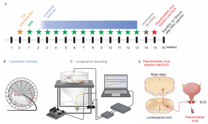
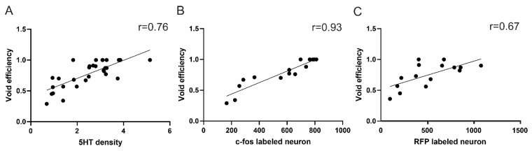
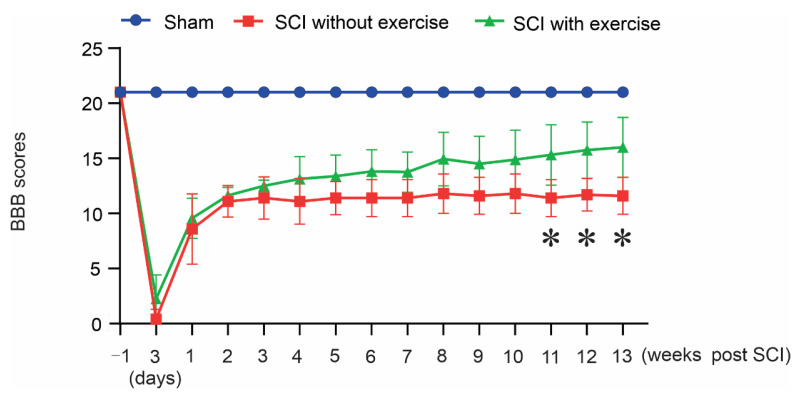
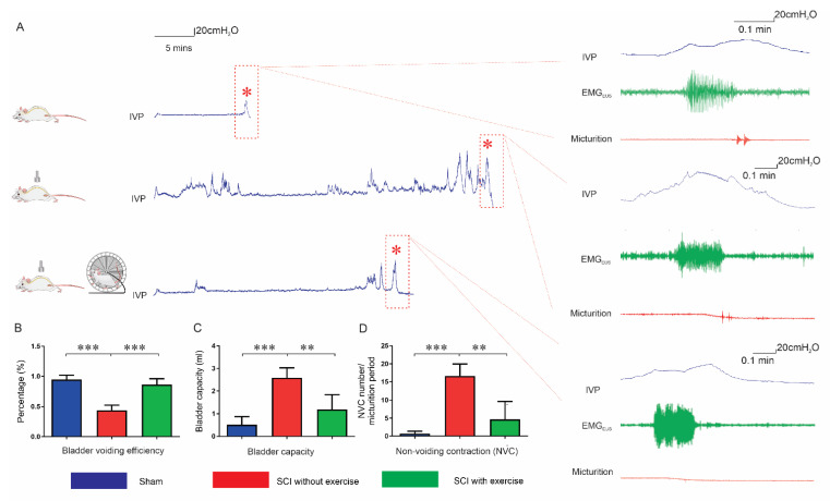
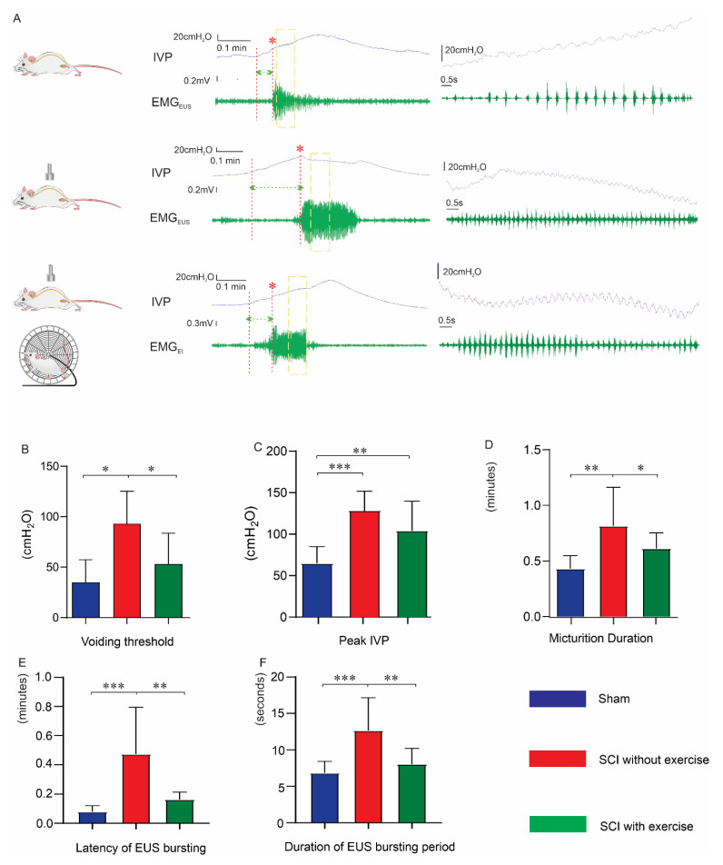
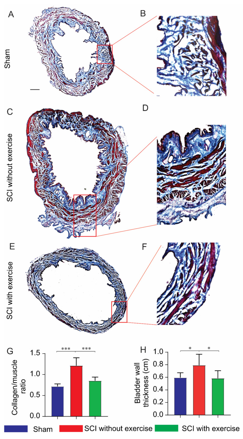
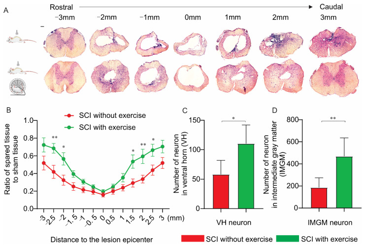
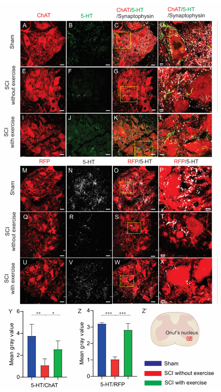
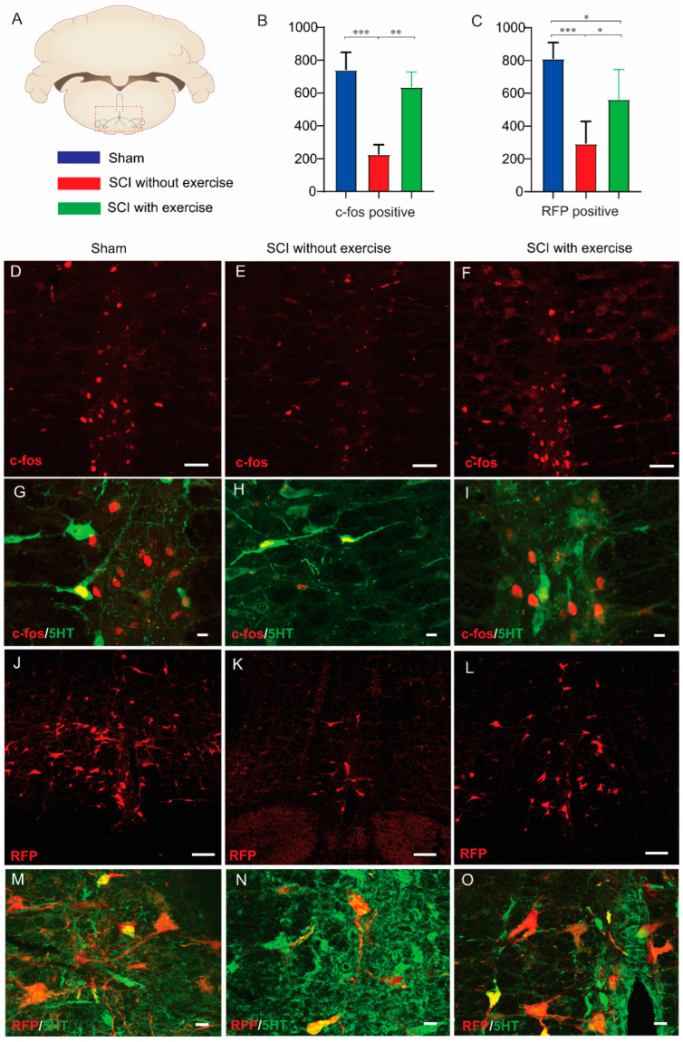
The recovery of lower-urinary-tract activity is a top priority for patients with spinal-cord injury. Historically, locomotor training improved micturition function in both patients with spinal cord injury and animal models. We explore whether training augments such as the supraspinal control of the external urethral sphincter results in enhanced coordination in detrusor-sphincter activity. We implemented a clinically relevant contusive spinal-cord injury at the 12th thoracic level in rats and administered forced wheel running exercise for 11 weeks. Awake rats then underwent bladder cystometrogram and sphincter electromyography recordings to examine the micturition reflex. Subsequently, pseudorabies-virus-encoding red fluorescent protein was injected into the sphincter to trans-synaptically trace the supraspinal innervation of Onuf's motoneurons. Training in the injury group reduced the occurrence of bladder nonvoiding contractions, decreased the voiding threshold and peak intravesical pressure, and shortened the latency of sphincter bursting during voiding, leading to enhanced voiding efficiency. Histological analysis demonstrated that the training increased the extent of spared spinal-cord tissue around the epicenter of lesions. Compared to the group of injury without exercise, training elicited denser 5-hydroxytryptamine-positive axon terminals in the vicinity of Onuf's motoneurons in the cord; more pseudorabies virus-labeled or c-fos expressing neurons were detected in the brainstem, suggesting the enhanced supraspinal control of sphincter activity. Thus, locomotor training promotes tissue sparing and axon innervation of spinal motoneurons to improve voiding function following contusive spinal-cord injury.
下尿路活动的恢复是脊髓损伤患者的首要任务。历史上,运动训练改善了脊髓损伤患者和动物模型的排尿功能。我们探讨了训练是否增强了尿道外括约肌的上位中枢控制,从而增强了逼尿肌-括约肌活动的协调性。我们在大鼠第 12 胸椎水平实施了具有临床相关性的挫伤性脊髓损伤,并进行了 11 周的强制轮跑运动。然后,清醒的大鼠进行膀胱测压和括约肌肌电图记录,以检查排尿反射。随后,将伪狂犬病毒编码的红色荧光蛋白注射到括约肌中,以转突触追踪 Onuf 运动神经元的上位神经支配。损伤组的训练减少了膀胱非排空收缩的发生,降低了排空阈值和膀胱内压峰值,并缩短了排尿时括约肌爆发的潜伏期,从而提高了排空效率。组织学分析表明,训练增加了损伤中心周围脊髓组织的保留程度。与无运动训练的损伤组相比,训练在脊髓中 Onuf 运动神经元附近引起了更密集的 5-羟色胺阳性轴突末梢;在脑干中检测到更多的伪狂犬病毒标记或 c-fos 表达神经元,表明括约肌活动的上位中枢控制增强。因此,运动训练促进了脊髓运动神经元的组织保留和轴突支配,从而改善了挫伤性脊髓损伤后的排尿功能。