Center for Image Sciences, Department of Radiology, University Medical Centre Utrecht, Utrecht.
Danish Research Centre for Magnetic Resonance, Centre for Functional and Diagnostic Imaging and Research, Copenhagen University Hospital Hvidovre, Hvidovre, Denmark.
NMR Biomed. 2022 Oct;35(10):e4771. doi: 10.1002/nbm.4771. Epub 2022 May 26.
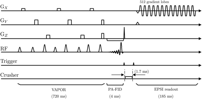
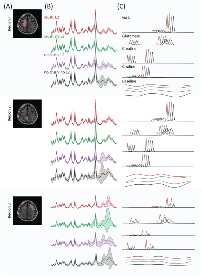
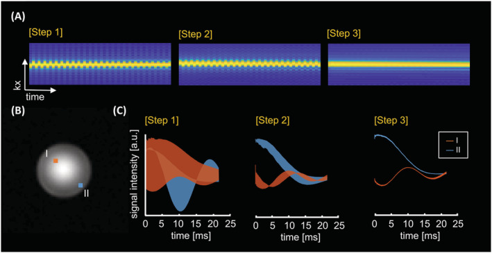
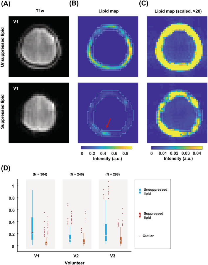
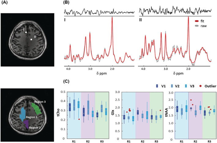
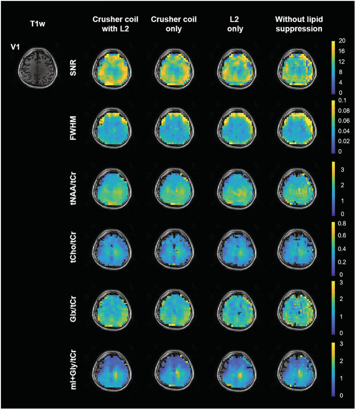
The increased signal-to-noise ratio (SNR) and chemical shift dispersion at high magnetic fields (≥7 T) have enabled neuro-metabolic imaging at high spatial resolutions. To avoid very long acquisition times with conventional magnetic resonance spectroscopic imaging (MRSI) phase-encoding schemes, solutions such as pulse-acquire or free induction decay (FID) sequences with short repetition time and inner volume selection methods with acceleration (echo-planar spectroscopic imaging [EPSI]), have been proposed. With the inner volume selection methods, limited spatial coverage of the brain and long echo times may still impede clinical implementation. FID-MRSI sequences benefit from a short echo time and have a high SNR per time unit; however, contamination from strong extra-cranial lipid signals remains a problem that can hinder correct metabolite quantification. L2-regularization can be applied to remove lipid signals in cases with high spatial resolution and accurate prior knowledge. In this work, we developed an accelerated two-dimensional (2D) FID-MRSI sequence using an echo-planar readout and investigated the performance of lipid suppression by L2-regularization, an external crusher coil, and the combination of these two methods to compare the resulting spectral quality in three subjects. The reduction factor of lipid suppression using the crusher coil alone varies from 2 to 7 in the lipid region of the brain boundary. For the combination of the two methods, the average lipid area inside the brain was reduced by 2% to 38% compared with that of unsuppressed lipids, depending on the subject's region of interest. 2D FID-EPSI with external lipid crushing and L2-regularization provides high in-plane coverage and is suitable for investigating brain metabolite distributions at high fields.
高磁场(≥7T)下信噪比(SNR)和化学位移弥散度的提高使神经代谢成像能够达到更高的空间分辨率。为了避免传统磁共振波谱成像(MRSI)相位编码方案的采集时间过长,可以采用脉冲采集或自由感应衰减(FID)序列,其重复时间短,并且具有加速的内体积选择方法(回波平面波谱成像[EPSI])。采用内体积选择方法,脑的空间覆盖范围有限且回波时间较长,这仍然会阻碍临床应用。FID-MRSI 序列具有短回波时间和单位时间高 SNR 的优点;然而,来自颅外强脂质信号的污染仍然是一个问题,可能会阻碍代谢物的正确定量。L2 正则化可用于在高空间分辨率和准确先验知识的情况下去除脂质信号。在这项工作中,我们使用回波平面读出方法开发了一种加速二维(2D)FID-MRSI 序列,并研究了 L2 正则化、外部碎裂线圈和这两种方法相结合对脂质抑制性能的影响,以比较三种受试者的光谱质量。单独使用碎裂线圈的脂质抑制的降低因子在脑边界的脂质区域内从 2 变化到 7。对于两种方法的组合,与未抑制的脂质相比,大脑内的平均脂质区域减少了 2%到 38%,具体取决于受试者的感兴趣区域。具有外部脂质碎裂和 L2 正则化的 2D FID-EPSI 提供了高的平面内覆盖范围,适合在高场研究大脑代谢物分布。