Paediatric Orthopaedics, Department of Trauma, Orthopaedic and Plastic Surgery, University Medical Center Goettingen, 37075, Goettingen, Germany.
Department of Trauma Surgery, Orthopaedic and Plastic Surgery, University Medical Center Goettingen, 37075, Goettingen, Germany.
Osteoporos Int. 2022 Sep;33(9):2011-2018. doi: 10.1007/s00198-022-06416-9. Epub 2022 May 18.
Duchenne muscular dystrophy is a progressive disease usually associated with loss of ambulation and progressive scoliosis. Immobilisation and glucocorticoid treatment are predisposing factors for reduced bone mineral density (BMD). Analysis of quantitative computed tomography revealed low BMD in thoracic and lumbar vertebrae in comparison to age- and sex-matched healthy controls.
Evaluation of vertebral bone mineral density (BMD) in Duchenne Muscular Dystrophy (DMD) adolescents with untreated advanced scoliosis and comparison with the BMD values of healthy age-matched controls, based on quantitative computer tomography.
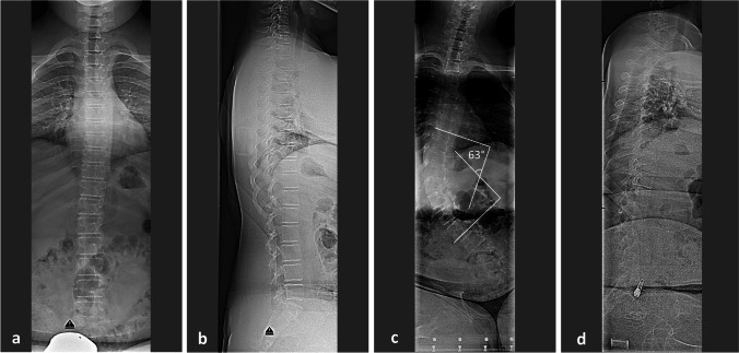
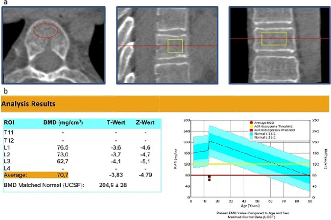
Thirty-seven DMD adolescents (age 15.6 ± 2.5 years) with spinal deformity were evaluated clinically and radiologically prior to definite spinal fusion and compared to 31 male and age-matched healthy individuals (age 15.7 ± 2.3 years). Data related to previous medical treatment, physiotherapy and ambulatory status was also analysed. Scoliotic curves were measured on plain sitting radiographs of the spine. The BMD Z-scores of the thoracic and lumbar vertebrae were calculated with QCTpro® (Mindways Software Inc., USA), based on data sets of preoperative, phantom pre-calibrated spinal computed tomography scans.
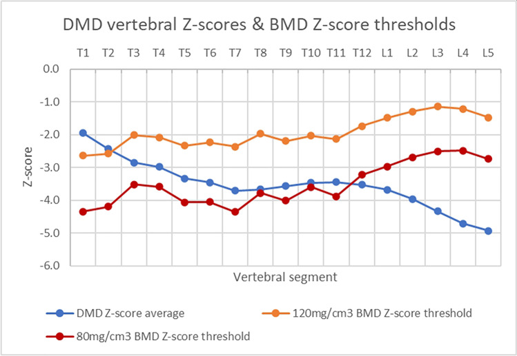
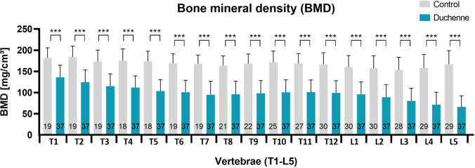
A statistically significant lower BMD could be found in DMD adolescents, when compared to healthy controls, showing an average value for the lumbar spine of 80.5 ± 30.5 mg/cm. Z-scores deteriorated from the upper thoracic towards the lower lumbar vertebrae. All but the uppermost thoracic vertebrae had reduced BMD values, with the thoracolumbar and lumbar region demonstrating the lowest BMD. No significant correlation was observed between BMD and the severity of the scoliotic curve, previous glucocorticoid treatment, cardiovascular impairment, vitamin D supplementation, non-invasive ventilation or physiotherapy.
DMD adolescents with scoliosis have strongly reduced BMD Z-scores, especially in the lumbar spine in comparison to healthy controls. These findings support the implementation of a standardised screening and treatment protocol. Level of evidence/clinical relevance: therapeutic level III.
评估未经治疗的进展性脊柱侧凸的杜氏肌营养不良症(DMD)青少年的椎体骨密度(BMD),并与健康年龄匹配对照者的 BMD 值进行比较,方法:对 37 例脊柱畸形的 DMD 青少年(年龄 15.6 ± 2.5 岁)进行临床和影像学评估,然后进行明确的脊柱融合,并与 31 名男性和年龄匹配的健康个体(年龄 15.7 ± 2.3 岁)进行比较。还分析了与以前的药物治疗、物理治疗和活动状态有关的数据。脊柱侧凸曲线在脊柱的普通坐姿射线照片上进行测量。根据术前、预制校准的脊柱计算机断层扫描数据集,使用 QCTpro®(Mindways Software Inc.,美国)计算胸椎和腰椎的 BMD Z 评分。结果:与健康对照组相比,DMD 青少年的 BMD 明显降低,腰椎平均为 80.5 ± 30.5mg/cm。Z 评分从胸椎上部向腰椎下部恶化。除了最上面的胸椎外,所有的胸椎都有降低的 BMD 值,胸腰椎和腰椎区域的 BMD 值最低。BMD 与脊柱侧凸曲线的严重程度、以前的糖皮质激素治疗、心血管损伤、维生素 D 补充、无创通气或物理治疗之间没有显著相关性。结论:患有脊柱侧凸的 DMD 青少年的 BMD Z 评分明显降低,与健康对照组相比,尤其是在腰椎。这些发现支持实施标准化的筛查和治疗方案。证据水平/临床相关性:治疗水平 III。