Edwin L. Steele Laboratories, Department of Radiation Oncology, Massachusetts General Hospital, Harvard Medical School, Boston, Massachusetts.
Department of Chemical Engineering, Massachusetts Institute of Technology, Cambridge, Massachusetts.
Clin Cancer Res. 2022 Jul 15;28(14):3076-3090. doi: 10.1158/1078-0432.CCR-22-0486.
The abnormal function of tumor blood vessels causes tissue hypoxia, promoting disease progression and treatment resistance. Although tumor microenvironment normalization strategies can alleviate hypoxia globally, how local oxygen levels change is not known because of the inability to longitudinally assess vascular and interstitial oxygen in tumors with sufficient resolution. Understanding the spatial and temporal heterogeneity should help improve the outcome of various normalization strategies.
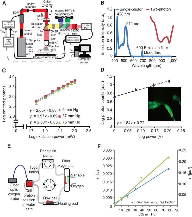
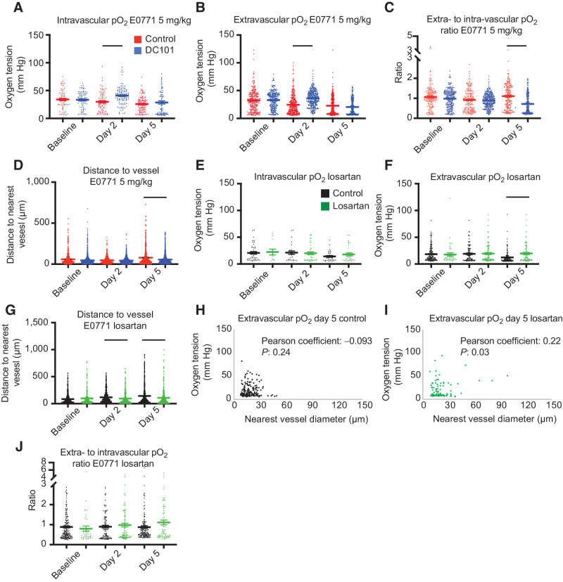
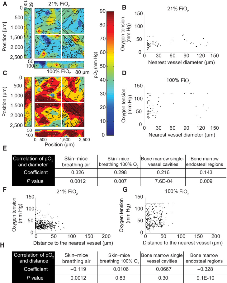
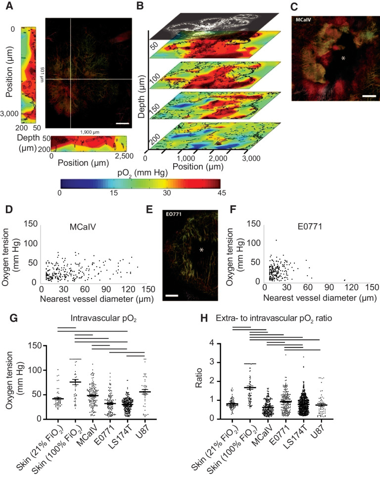
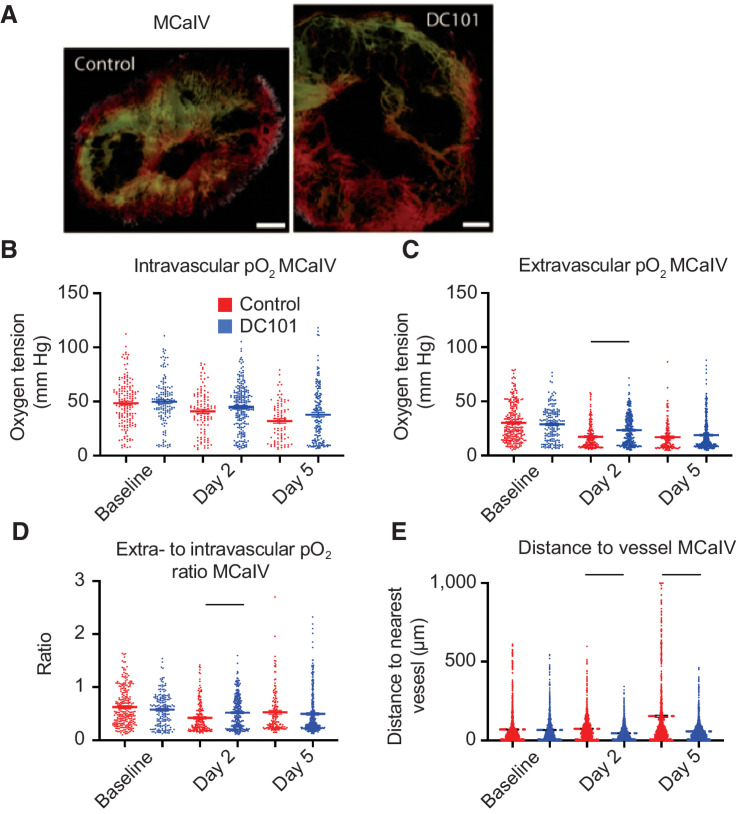
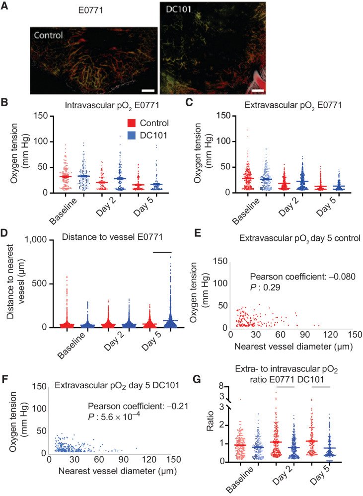
We developed a multiphoton phosphorescence quenching microscopy system using a low-molecular-weight palladium porphyrin probe to measure perfused vessels, oxygen tension, and their spatial correlations in vivo in mouse skin, bone marrow, and four different tumor models. Further, we measured the temporal and spatial changes in oxygen and vessel perfusion in tumors in response to an anti-VEGFR2 antibody (DC101) and an angiotensin-receptor blocker (losartan).
We found that vessel function was highly dependent on tumor type. Although some tumors had vessels with greater oxygen-carrying ability than those of normal skin, most tumors had inefficient vessels. Further, intervessel heterogeneity in tumors is associated with heterogeneous response to DC101 and losartan. Using both vascular and stromal normalizing agents, we show that spatial heterogeneity in oxygen levels persists, even with reductions in mean extravascular hypoxia.
High-resolution spatial and temporal responses of tumor vessels to two agents known to improve vascular perfusion globally reveal spatially heterogeneous changes in vessel structure and function. These dynamic vascular changes should be considered in optimizing the dose and schedule of vascular and stromal normalizing strategies to improve the therapeutic outcome.
肿瘤血管的异常功能导致组织缺氧,从而促进疾病的进展和治疗耐药性。尽管肿瘤微环境正常化策略可以全局缓解缺氧,但由于无法以足够的分辨率纵向评估肿瘤中的血管和间质氧,因此不知道局部氧水平的变化情况。了解空间和时间异质性应该有助于改善各种正常化策略的结果。
我们开发了一种多光子磷光猝灭显微镜系统,使用低分子量钯卟啉探针来测量体内小鼠皮肤、骨髓和四种不同肿瘤模型中的灌注血管、氧张力及其空间相关性。此外,我们还测量了肿瘤中氧和血管灌注对抗 VEGFR2 抗体(DC101)和血管紧张素受体阻滞剂(氯沙坦)的时空变化。
我们发现血管功能高度依赖于肿瘤类型。尽管一些肿瘤的血管携氧能力大于正常皮肤的血管,但大多数肿瘤的血管效率较低。此外,肿瘤内血管间的异质性与 DC101 和氯沙坦的异质反应相关。我们使用血管和基质正常化剂表明,即使在血管外缺氧的平均水平降低的情况下,氧水平的空间异质性仍然存在。
两种已知可改善血管灌注的药物对肿瘤血管的高分辨率时空反应揭示了血管结构和功能的空间异质性变化。在优化血管和基质正常化策略的剂量和方案以改善治疗结果时,应考虑这些动态血管变化。