Department of Anatomy and Embryology, Faculty of Veterinary Medicine, Sohag University, Sohag, 82524, Egypt.
Department of Anatomy and Embryology, Faculty of Veterinary Medicine, South Valley University, Qena, 83523, Egypt.
Sci Rep. 2022 May 18;12(1):8334. doi: 10.1038/s41598-022-12243-z.
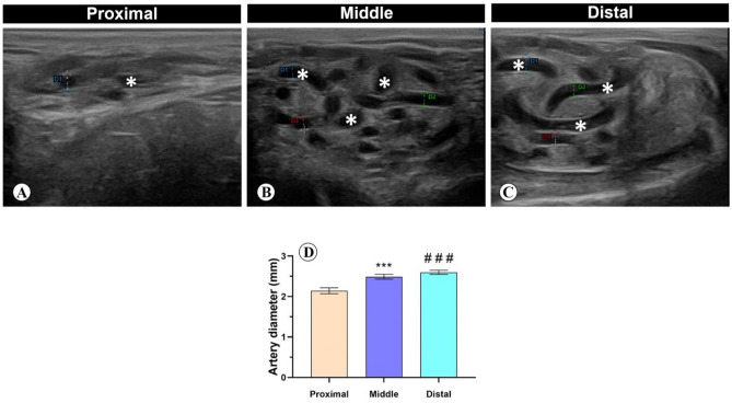
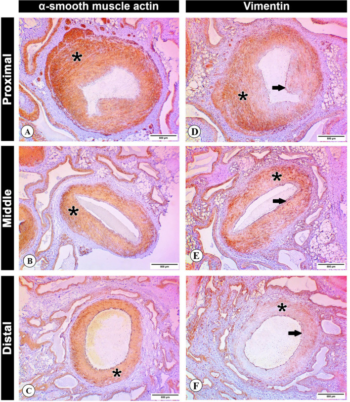
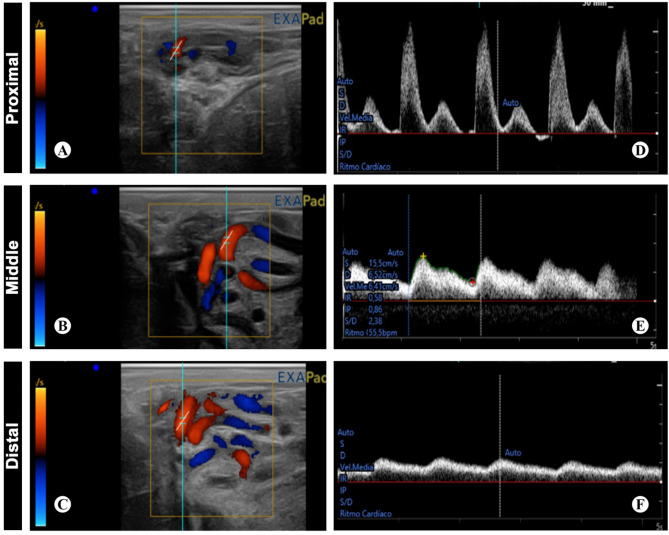
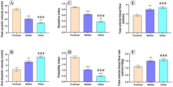
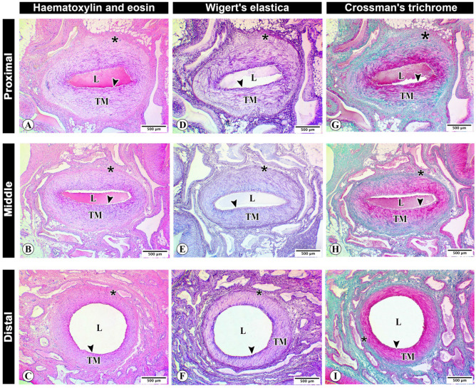
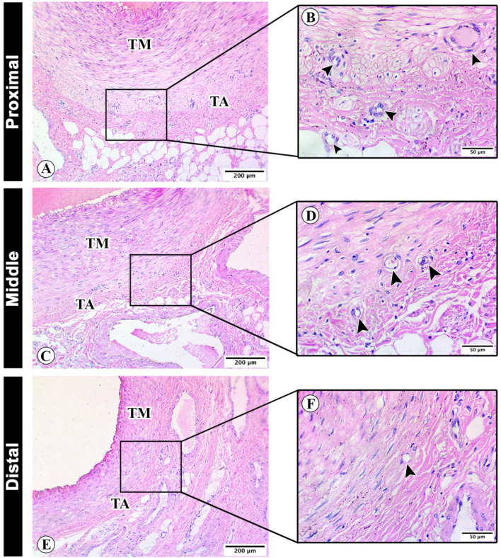
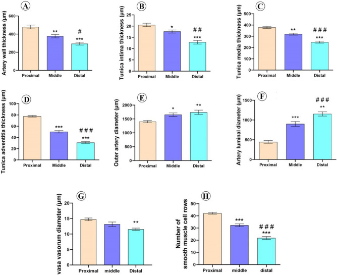
To fully understand the histological, morphometrical and heamodynamic variations of different supratesticular artery regions, 20 mature and healthy Assaf rams were examined through ultrasound and morphological studies. The testicular artery images of the spermatic cord as shown by B-mode analysis indicated a tortuous pattern along its course toward the testis, although it tends to be less tortuous close to the inguinal ring. Doppler velocimetric values showed a progressive decline in flow velocity, in addition to pulsatility and vessel resistivity when entering the testis, where there were significant differences in the Doppler indices and velocities among the different regions. The peak systolic velocity, pulsatility index and resistive index were higher in the proximal supratesticular artery region, followed by middle and distal ones, while the end diastolic velocity was higher in the distal supratesticular region. The total arterial blood flow and total arterial blood flow rate reported a progressive and significant increase along the testicular cord until entering the testis. Histological examination revealed presence of vasa vasorum in the tunica adventitia, with their diameter is higher in the proximal supratesticular zone than middle and distal ones. Morphometrically, the thickness of the supratesticular artery wall showed a significant decline downward toward the testis; meanwhile, the outer arterial diameter and inner luminal diameter displayed a significant increase distally. The expression of alpha smooth muscle actin and vimentin was higher in the tunica media of the proximal supratesticular artery zone than in middle and distal ones.
为了充分了解不同精索外动脉区域的组织学、形态计量学和血液动力学变化,通过超声和形态学研究检查了 20 只成熟健康的阿萨夫公羊。B 型分析显示精索睾丸动脉图像在向睾丸延伸的过程中呈扭曲模式,尽管在靠近腹股沟环处它的扭曲程度较小。多普勒速度值显示在进入睾丸时,血流速度、脉动指数和血管阻力逐渐下降,在不同区域之间存在明显的多普勒指数和速度差异。收缩期峰值速度、脉动指数和阻力指数在精索外动脉近端区域较高,其次是中部和远端区域,而舒张末期速度在精索外动脉远端区域较高。总动脉血流量和总动脉血流速率沿着睾丸索呈逐渐显著增加,直到进入睾丸。组织学检查显示在鞘膜外膜中存在血管周细胞,其直径在精索外动脉近端区域高于中部和远端区域。形态计量学上,精索外动脉壁的厚度向睾丸方向呈显著下降趋势;同时,外动脉直径和内腔直径在远端呈显著增加。在精索外动脉近端区域的中膜中,α平滑肌肌动蛋白和波形蛋白的表达高于中部和远端区域。