Pan Xian, Wang Han-Lu, Lin Shi-Ming, Lin Jia-Li, Ruan Dan-Dan, Zhang Jian-Hui, Chen Ting, Luo Jie-Wei, Fang Zhu-Ting
Fujian Provincial Hospital, Shengli Clinical Medical College of Fujian Medical University, Fuzhou, China.
Department of Interventional Radiology, Fujian Provincial Hospital, Fuzhou, China.
Front Oncol. 2022 May 2;12:892943. doi: 10.3389/fonc.2022.892943. eCollection 2022.
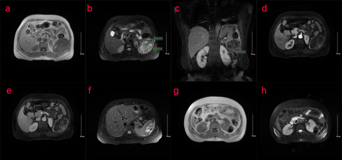
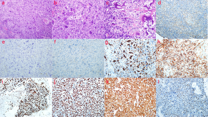
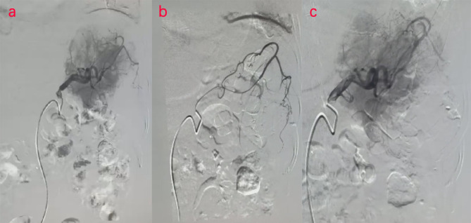
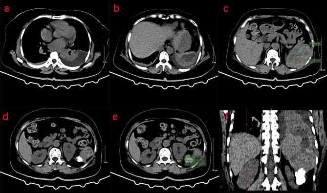
Extraskeletal osteosarcoma is a rare malignant soft-tissue sarcoma that is difficult to diagnose. Surgery is a common treatment, although chemotherapy and radiotherapy are also used. Patients at risk of bleeding can undergo embolization combined with resection. The occurrence of primary splenic extraskeletal osteosarcoma in humans does not seem to have been reported in the literature. A 50-year-old woman who complained of pain in the left upper abdomen for 1 day was initially diagnosed with "splenic hemangioma with a high possibility of rupture and bleeding" and urgently underwent digital subtraction angiography, combined with splenic arteriography and embolization. Abdominal pain worsened 2 days postoperatively, with a hemoglobin level of 106.0 g/L. Consequently, emergency laparotomy combined with splenectomy was performed. The clinical and imaging features, pathological diagnosis, and embolization treatment of this case were analyzed retrospectively. CT of the upper abdomen revealed splenomegaly, an irregular low-density shadow in the spleen, and a flake-like calcification in the lateral margin of the left kidney. Nuclear MRI of the upper abdomen showed splenomegaly and a mass (approximately 8.4 cm × 5.7 cm × 6.3 cm) below the spleen with clear boundaries-this exhibited an uneven signal, which was slightly low in T1-weighted imaging (T1WI) and slightly high in T2-weighted imaging (T2WI). Several small cystic lesions or cystic cavities were observed in the mass, which exhibited a longer T2 signal. During the enhanced scan, the signal of the lesion showed progressive enhancement, and the enhancement range increased in the delayed phase scan, as well as a hematoma below the spleen capsule and calcification below the lesion (nodular T1WI/T2WI hypointense, approximately 3.3 cm × 3.6 cm). Postoperative biopsy pathology showed splenic soft tissue tumor: at low magnification, the multinucleated giant cells were scattered; at medium magnification, osteoclast-like multinucleated giant cells were observed; and at high magnification, lace- or grid-like tumor osteogenesis was detected. Immunohistochemistry showed that the expression of CD31, CD34, F8, s-100, desmin, SMA, and CD99 was negative, whereas the expression of β-catenin, BCL-2, SATB-2, and P16 was positive. CD68 and MDM-2 showed low expression, while 50% of the cells were positive for Ki-67 expression. No abnormal concentration of radioactivity was found on the bone scan with Tc-MDP after the operation, further ruling out the occurrence of other bone tumors. The patient was diagnosed with primary extraskeletal osteosarcoma. It is necessary for multidisciplinary teams to diagnose malignant extraskeletal osteosarcomas.
骨外骨肉瘤是一种罕见的恶性软组织肉瘤,难以诊断。手术是常见的治疗方法,不过也会使用化疗和放疗。有出血风险的患者可接受栓塞联合切除术。原发性脾脏骨外骨肉瘤在人类中的发生似乎尚未见文献报道。一名50岁女性因左上腹疼痛1天就诊,最初被诊断为“高度疑似破裂出血的脾脏血管瘤”,并紧急接受了数字减影血管造影,联合脾动脉造影和栓塞术。术后2天腹痛加重,血红蛋白水平为106.0 g/L。因此,进行了急诊剖腹探查联合脾切除术。对该病例的临床及影像学特征、病理诊断及栓塞治疗进行回顾性分析。上腹部CT显示脾肿大,脾内有不规则低密度影,左肾外侧缘有片状钙化。上腹部核磁共振成像显示脾肿大,脾脏下方有一肿块(约8.4 cm×5.7 cm×6.3 cm),边界清晰,信号不均匀,在T1加权成像(T1WI)上略低,在T2加权成像(T2WI)上略高。肿块内可见多个小囊性病变或囊腔,呈长T2信号。增强扫描时,病变信号呈渐进性强化,延迟期扫描强化范围增大,脾包膜下有血肿,病变下方有钙化(结节状T1WI/T2WI低信号,约3.3 cm×3.6 cm)。术后活检病理显示脾脏软组织肿瘤:低倍镜下可见多核巨细胞散在分布;中倍镜下可见破骨细胞样多核巨细胞;高倍镜下可见花边状或网格状肿瘤骨形成。免疫组化显示CD31、CD34、F8、s-100、结蛋白、平滑肌肌动蛋白和CD99表达均为阴性,而β-连环蛋白、BCL-2、SATB-2和P16表达为阳性。CD68和MDM-2呈低表达,Ki-67表达阳性的细胞占50%。术后用锝-亚甲基二膦酸盐进行骨扫描未发现放射性异常浓聚,进一步排除了其他骨肿瘤的发生。该患者被诊断为原发性骨外骨肉瘤。多学科团队对恶性骨外骨肉瘤进行诊断很有必要。