Department of Biomedical Sciences, University of Missouri, Columbia, Missouri.
Dalton Cardiovascular Research Center, University of Missouri, Columbia, Missouri.
J Neurophysiol. 2022 Jul 1;128(1):28-39. doi: 10.1152/jn.00102.2022. Epub 2022 Jun 1.
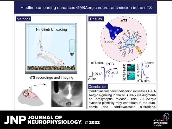
The nucleus tractus solitarii (nTS) is the major integrative brainstem region for autonomic modulation and processing of cardiovascular reflexes. GABA and glutamate are the main inhibitory and excitatory neurotransmitters, respectively, within this nucleus. Alterations in the GABA-glutamate regulation in the nTS are related to numerous cardiovascular comorbidities. Bedridden individuals and people exposed to microgravity exhibit dysautonomia and cardiovascular deconditioning that are mimicked in the hindlimb unloading (HU) rat model. We have previously shown in the nTS that HU increases glutamatergic neurotransmission yet decreases neuronal excitability. In this study, we investigated the effects of HU on nTS GABAergic neurotransmission. We hypothesized that HU potentiates GABA signaling via increased GABAergic release and postsynaptic GABA receptor expression. Following HU or control postural exposure, GABAergic neurotransmission was assessed using whole cell patch clamp whereas the magnitude of GABA release was evaluated via an intensity-based GABA sensing fluorescence reporter (iGABASnFR). In response to GABA interneuron stimulation, the evoked inhibitory postsynaptic current (nTS-IPSC) amplitude and area, as well as iGABASnFR fluorescence, were greater in HU than in control. HU also elevated the frequency but not the amplitude of spontaneous miniature IPSCs. Picoapplication of GABA produced similar postsynaptic current responses in nTS neurons of HU and control. Moreover, HU did not alter GABA receptor α1 subunit expression, indicating minimal alterations in postsynaptic membrane receptor expression. These results indicate that HU increases GABAergic signaling in the nTS likely via augmented release of GABA from presynaptic terminals. Altogether, our data indicate GABA plasticity contributes to the autonomic and cardiovascular alterations following cardiovascular deconditioning (CVD). Gravity influences distribution of blood volume and autonomic function. Microgravity and prolonged bed rest induce cardiovascular deconditioning (CVD). We used hindlimb unloading (HU), a rat analog for bed rest, to investigate CVD-induced neuroplasticity in the brainstem. Our data demonstrate that HU increases GABA modulation of nucleus tractus solitarii (nTS) neurons via presynaptic plasticity. Given the importance of nTS in integrating cardiovascular reflexes, this study provides new evidence on the central mechanisms behind CVD following HU.
孤束核(nTS)是自主调节和心血管反射处理的主要整合脑干区域。GABA 和谷氨酸分别是该核内的主要抑制性和兴奋性神经递质。nTS 中的 GABA-谷氨酸调节的改变与许多心血管合并症有关。卧床不起的个体和暴露于微重力的个体表现出自主神经功能障碍和心血管适应不良,这些在下肢悬吊(HU)大鼠模型中得到了模拟。我们之前在 nTS 中表明,HU 增加了谷氨酸能神经传递,但降低了神经元兴奋性。在这项研究中,我们研究了 HU 对 nTS GABA 能神经传递的影响。我们假设 HU 通过增加 GABA 能释放和突触后 GABA 受体表达来增强 GABA 信号。在 HU 或对照姿势暴露后,使用全细胞贴片钳评估 GABA 能神经传递,而通过基于强度的 GABA 感应荧光报告物(iGABASnFR)评估 GABA 释放的幅度。响应 GABA 中间神经元刺激,HU 中的诱发抑制性突触后电流(nTS-IPSC)幅度和面积以及 iGABASnFR 荧光均大于对照。HU 还增加了自发性微小 IPSC 的频率但不增加幅度。GABA 的皮科应用在 HU 和对照的 nTS 神经元中产生类似的突触后电流反应。此外,HU 并未改变 GABA 受体 α1 亚基的表达,表明突触后膜受体表达的变化很小。这些结果表明,HU 通过增加来自突触前末端的 GABA 释放来增加 nTS 中的 GABA 能信号。总的来说,我们的数据表明 GABA 可塑性有助于心血管适应不良(CVD)后自主和心血管的改变。重力影响血液量和自主功能的分布。微重力和长时间卧床会导致心血管适应不良(CVD)。我们使用下肢悬吊(HU),一种卧床休息的大鼠模拟物,研究了脑干中 CVD 诱导的神经可塑性。我们的数据表明,HU 通过突触前可塑性增加了孤束核(nTS)神经元的 GABA 调节。鉴于 nTS 在整合心血管反射中的重要性,这项研究为 HU 后 CVD 的中枢机制提供了新的证据。