Tsugawa Anson J, Arzi Boaz, Vapniarsky Natalia, Verstraete Frank J M
School of Veterinary Medicine, William R. Pritchard Veterinary Medical Teaching Hospital, University of California, Davis, Davis, CA, United States.
Department of Surgical and Radiological Sciences, School of Veterinary Medicine, University of California, Davis, Davis, CA, United States.
Front Vet Sci. 2022 May 11;9:900031. doi: 10.3389/fvets.2022.900031. eCollection 2022.
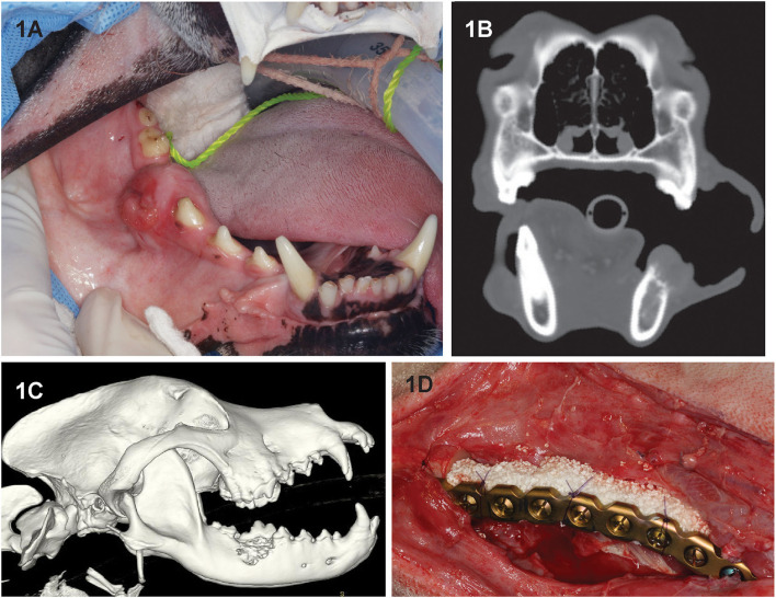
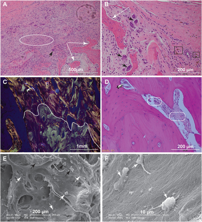
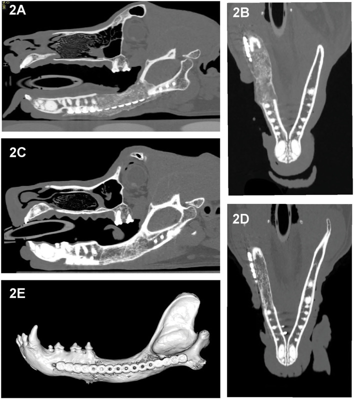
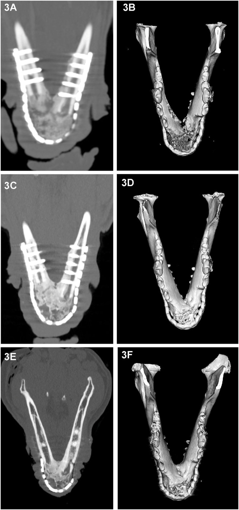
The successful excision of a locally invasive tumor such as canine acanthomatous ameloblastoma (CAA) typically results in a mandibular contour-derforming, critical-size defect that alters the jaw kinematics, and may affect the patient's quality of life. In this case series, we describe our experience using the regenerative approach of a titanium locking plate and compression resistant matrix infused with rhBMP-2 for the immediate or delayed reconstruction following mandibulectomy for the excision of mandibular CAA in 11 dogs. Surgical planning included computed tomography (CT), with and without contrast, in all cases, and 3D-printed models in four cases. Tumor-free surgical margins were achieved in all dogs. Clinical and diagnostic imaging follow-up (mean, 23.1 months) were performed in-person (11 cases) and with CT/cone-beam computed tomography in most cases, with standard radiography (3 cases) and telemedicine being utilized in 5 cases. At 2 weeks postoperatively, hard tissue was palpable at the defect. Follow-up imaging at 1 month postoperatively revealed evidence of bridging new bone with a heterogeneous appearance, that remodeled over 3-6 months to bone of a similar size, shape and trabecular pattern as native bone. Histological evaluation of regenerated bone was available in two cases, and was supportive of our clinical and imaging findings of normal remodeled bone. Clinically, all dogs returned to a normal lifestyle, rapidly resumed eating and drinking, and exhibited normal occlusion. Complications included wound dehiscence in one dog and self-limiting exuberant bone formation in two dogs. Tumor regrowth, failure of the implant or fracture of the regenerated bone were not observed. We conclude that the mandibular reconstruction using a regenerative approach is safe, feasible, and results in restoration of mandibular contour in dogs following segmental and bilateral rostral mandibulectomy for benign but invasive oral tumors such as CAA.
成功切除局部侵袭性肿瘤,如犬棘皮瘤性成釉细胞瘤(CAA),通常会导致下颌轮廓变形、临界尺寸缺损,改变颌骨运动学,并可能影响患者的生活质量。在本病例系列中,我们描述了我们使用钛锁定板和注入重组人骨形态发生蛋白-2(rhBMP-2)的抗压基质的再生方法,对11只犬切除下颌CAA后进行下颌骨切除术后即刻或延迟重建的经验。所有病例的手术规划均包括有或无对比剂的计算机断层扫描(CT),4例使用了3D打印模型。所有犬均实现了无瘤手术切缘。临床和诊断性影像随访(平均23.1个月)通过亲自检查(11例)进行,大多数病例采用CT/锥形束计算机断层扫描,3例采用标准X线摄影,5例采用远程医疗。术后2周,在缺损处可触及硬组织。术后1个月的随访影像显示有桥接新骨的迹象,其外观不均匀,在3至6个月内重塑为与天然骨大小、形状和小梁模式相似的骨。两例对再生骨进行了组织学评估,支持了我们对正常重塑骨的临床和影像结果。临床上,所有犬都恢复了正常生活方式,迅速恢复饮食,并表现出正常咬合。并发症包括1只犬伤口裂开和2只犬自限性骨过度生长。未观察到肿瘤复发、植入物失败或再生骨骨折。我们得出结论,对于良性但侵袭性的口腔肿瘤如CAA,采用再生方法进行下颌骨重建是安全、可行的,并且可以恢复犬下颌骨切除术后的下颌轮廓。