• 文献检索
  • 文档翻译
  • 深度研究
  • 学术资讯
  • Suppr Zotero 插件Zotero 插件
  • 邀请有礼
  • 套餐&价格
  • 历史记录
应用&插件
Suppr Zotero 插件Zotero 插件浏览器插件Mac 客户端Windows 客户端微信小程序
定价
高级版会员购买积分包购买API积分包
服务
文献检索文档翻译深度研究API 文档MCP 服务
关于我们
关于 Suppr公司介绍联系我们用户协议隐私条款
关注我们

Suppr 超能文献

核心技术专利:CN118964589B侵权必究
粤ICP备2023148730 号-1Suppr @ 2026

文献检索

告别复杂PubMed语法,用中文像聊天一样搜索,搜遍4000万医学文献。AI智能推荐,让科研检索更轻松。

立即免费搜索

文件翻译

保留排版,准确专业,支持PDF/Word/PPT等文件格式,支持 12+语言互译。

免费翻译文档

深度研究

AI帮你快速写综述,25分钟生成高质量综述,智能提取关键信息,辅助科研写作。

立即免费体验

对比增强乳腺摄影在筛查召回评估中的应用:一项两中心研究。

Contrast-enhanced mammography for the assessment of screening recalls: a two-centre study.

机构信息

Department of Biomedical Sciences for Health, Università degli Studi di Milano, Via Luigi Mangiagalli 31, 20133, Milano, Italy.

Unit of Radiology, IRCCS Policlinico San Donato, Via Rodolfo Morandi 30, 20097, San Donato Milanese, Italy.

出版信息

Eur Radiol. 2022 Nov;32(11):7388-7399. doi: 10.1007/s00330-022-08868-3. Epub 2022 Jun 1.

DOI:10.1007/s00330-022-08868-3
PMID:35648209
原文链接:https://pmc.ncbi.nlm.nih.gov/articles/PMC9668944/
Abstract

OBJECTIVES

To evaluate the potential of contrast-enhanced mammography (CEM) for reducing the biopsy rate of screening recalls.

METHODS

Recalled women were prospectively enrolled to undergo CEM alongside standard assessment (SA) through additional views, tomosynthesis, and/or ultrasound. Exclusion criteria were symptoms, implants, allergy to contrast agents, renal failure, and pregnancy. SA and CEM were independently evaluated by one of six radiologists, who recommended biopsy or 2-year follow-up. Biopsy rates according to SA or recombined CEM (rCEM) were compared with the McNemar's test. Diagnostic performance was calculated considering lesions with available final histopathology.

RESULTS

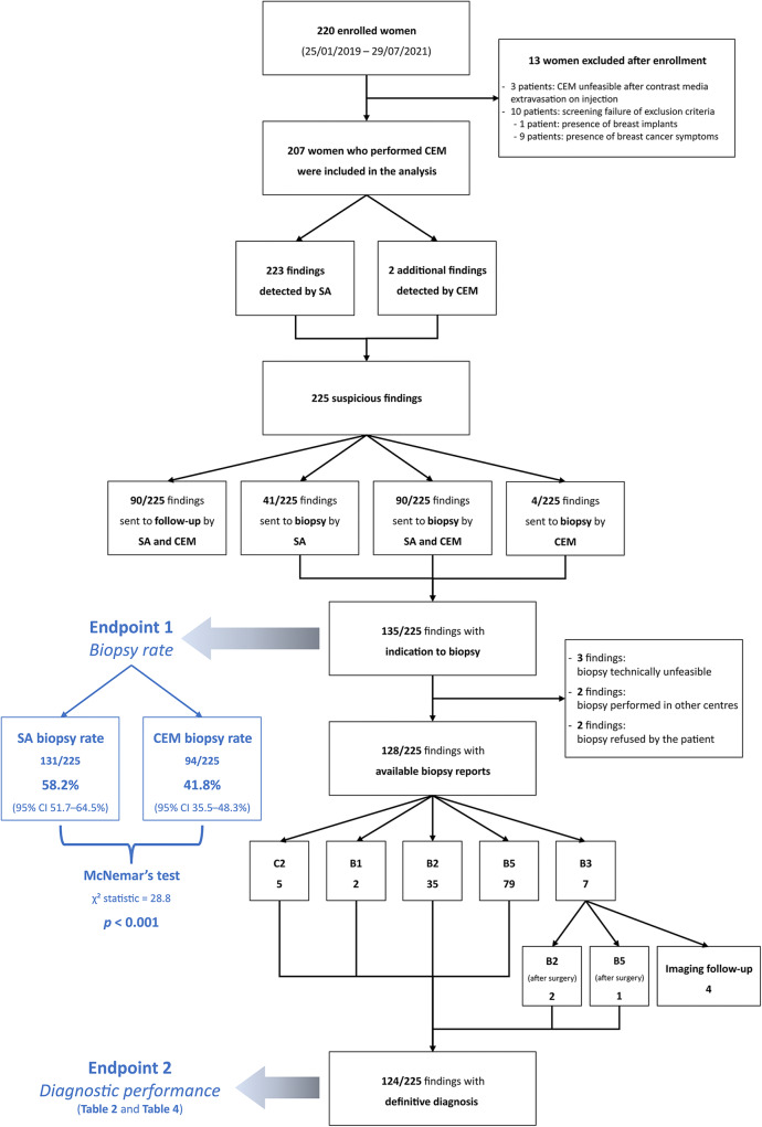
Between January 2019 and July 2021, 220 women were enrolled, 207 of them (median age 56.6 years) with 225 suspicious findings analysed. Three of 207 patients (1.4%) developed mild self-limiting adverse reactions to iodinated contrast agent. Overall, 135/225 findings were referred for biopsy, 90/225 by both SA and rCEM, 41/225 by SA alone and 4/225 by rCEM alone (2/4 being one DCIS and one invasive carcinoma). The rCEM biopsy rate (94/225, 41.8%, 95% CI 35.5-48.3%) was 16.4% lower (p < 0.001) than the SA biopsy rate (131/225, 58.2%, 95% CI 51.7-64.5%). Considering the 124/135 biopsies with final histopathology (44 benign, 80 malignant), rCEM showed a 93.8% sensitivity (95% CI 86.2-97.3%) and a 65.9% specificity (95% CI 51.1-78.1%), all 5 false negatives being ductal carcinoma in situ detectable as suspicious calcifications on low-energy images.

CONCLUSIONS

Compared to SA, the rCEM-based work-up would have avoided biopsy for 37/225 (16.4%) suspicious findings. Including low-energy images in interpretation provided optimal overall CEM sensitivity.

KEY POINTS

• The work-up of suspicious findings detected at mammographic breast cancer screening still leads to a high rate of unnecessary biopsies, involving between 2 and 6% of screened women. • In 207 recalled women with 225 suspicious findings, recombined images of contrast-enhanced mammography (CEM) showed a 93.8% sensitivity and a 65.9% specificity, all 5 false negatives being ductal carcinoma in situ detectable on low-energy images as suspicious calcifications. • CEM could represent an easily available one-stop shop option for the morphofunctional assessment of screening recalls, potentially reducing the biopsy rate by 16.4%.

摘要

目的

评估对比增强乳腺摄影(CEM)在降低筛查召回活检率方面的潜力。

方法

前瞻性纳入召回的女性,同时进行 CEM 检查和标准评估(SA),包括额外视图、断层合成和/或超声检查。排除标准为症状、植入物、造影剂过敏、肾衰竭和怀孕。由六名放射科医生中的一名独立评估 SA 和 CEM,并推荐活检或 2 年随访。比较 SA 或组合 CEM(rCEM)的活检率,并采用 McNemar 检验。考虑到具有可用最终组织病理学的病变,计算诊断性能。

结果

2019 年 1 月至 2021 年 7 月,共纳入 220 名女性,225 例可疑病变中 207 例(中位年龄 56.6 岁)进行了分析。207 名患者中有 3 名(1.4%)出现轻微的自限性碘造影剂不良反应。总体而言,225 例可疑病变中有 135 例被推荐活检,90 例同时通过 SA 和 rCEM 推荐,41 例仅通过 SA 推荐,4 例仅通过 rCEM 推荐(4 例中 2 例为 DCIS 和 1 例浸润性癌)。rCEM 活检率(94/225,41.8%,95%CI 35.5-48.3%)比 SA 活检率(131/225,58.2%,95%CI 51.7-64.5%)低 16.4%(p<0.001)。考虑到 124 例具有最终组织病理学的活检(44 例良性,80 例恶性),rCEM 显示出 93.8%的敏感性(95%CI 86.2-97.3%)和 65.9%的特异性(95%CI 51.1-78.1%),所有 5 例假阴性均为导管原位癌,在低能图像上可表现为可疑钙化。

结论

与 SA 相比,基于 rCEM 的检查可避免对 225 例可疑病变中的 37 例进行活检。在解释中纳入低能图像可提供最佳的整体 CEM 敏感性。

关键点

  • 在乳腺 X 线筛查的可疑病变检查中,仍有很高比例的不必要活检,涉及 2%至 6%的筛查女性。

  • 在 207 名有 225 例可疑病变的召回女性中,组合的 CEM 图像显示出 93.8%的敏感性和 65.9%的特异性,所有 5 例假阴性均为导管原位癌,在低能图像上可表现为可疑钙化。

  • CEM 可以作为筛查召回的形态功能评估的一种便捷的一站式选择,有可能将活检率降低 16.4%。

https://cdn.ncbi.nlm.nih.gov/pmc/blobs/dea0/9668944/df3570226459/330_2022_8868_Fig6_HTML.jpg
https://cdn.ncbi.nlm.nih.gov/pmc/blobs/dea0/9668944/971378f72824/330_2022_8868_Fig1_HTML.jpg
https://cdn.ncbi.nlm.nih.gov/pmc/blobs/dea0/9668944/d68200490b6d/330_2022_8868_Fig2_HTML.jpg
https://cdn.ncbi.nlm.nih.gov/pmc/blobs/dea0/9668944/ac9bc4b7e60e/330_2022_8868_Fig3_HTML.jpg
https://cdn.ncbi.nlm.nih.gov/pmc/blobs/dea0/9668944/16e94e2c53a9/330_2022_8868_Fig4_HTML.jpg
https://cdn.ncbi.nlm.nih.gov/pmc/blobs/dea0/9668944/1b108d3c9e2b/330_2022_8868_Fig5_HTML.jpg
https://cdn.ncbi.nlm.nih.gov/pmc/blobs/dea0/9668944/df3570226459/330_2022_8868_Fig6_HTML.jpg

相似文献

1
Contrast-enhanced mammography for the assessment of screening recalls: a two-centre study.对比增强乳腺摄影在筛查召回评估中的应用:一项两中心研究。
Eur Radiol. 2022 Nov;32(11):7388-7399. doi: 10.1007/s00330-022-08868-3. Epub 2022 Jun 1.
2
Failure of stereotactic core needle biopsy in women recalled for suspicious calcifications at screening mammography: frequency, causes, and final outcome in a multi-institutional, observational follow-up study.在筛查性乳房 X 光摄影中因可疑钙化而召回的女性中,立体定向核心针活检失败:多机构观察性随访研究中的频率、原因和最终结果。
Eur Radiol. 2022 Nov;32(11):7420-7429. doi: 10.1007/s00330-022-08806-3. Epub 2022 Apr 29.
3
The value of MRI contrast enhancement in biopsy decision of suspicious mammographic microcalcifications: a prospective multicenter study.MRI 对比增强在可疑乳腺微钙化活检决策中的价值:一项前瞻性多中心研究。
Eur Radiol. 2021 Mar;31(3):1718-1726. doi: 10.1007/s00330-020-07265-y. Epub 2020 Sep 16.
4
Contrast-Enhanced Mammography in Local Staging of Screen-Detected Breast Cancer.对比增强乳腺摄影在筛查性乳腺癌局部分期中的应用。
Ann Surg Oncol. 2024 Oct;31(10):6820-6830. doi: 10.1245/s10434-024-15848-y. Epub 2024 Jul 24.
5
Can positron emission mammography help to identify clinically significant breast cancer in women with suspicious calcifications on mammography?正电子发射乳腺断层成像能否帮助识别乳腺钼靶检查有可疑钙化的女性中的临床显著乳腺癌?
Eur Radiol. 2017 May;27(5):1893-1900. doi: 10.1007/s00330-016-4576-z. Epub 2016 Sep 2.
6
Stereotactic and sonographic large-core biopsy of nonpalpable breast lesions: results of the Radiologic Diagnostic Oncology Group V study.立体定向和超声引导下不可触及乳腺病变的粗针活检:放射诊断肿瘤学组V研究结果
Acad Radiol. 2004 Mar;11(3):293-308. doi: 10.1016/s1076-6332(03)00510-5.
7
Utility of MRI-Directed Contrast-Enhanced Mammography for Biopsy Planning for Suspicious MRI-Detected Breast Lesions.MRI 引导对比增强乳腺摄影在可疑 MRI 检出乳腺病变活检规划中的应用。
AJR Am J Roentgenol. 2023 Feb;220(2):202-211. doi: 10.2214/AJR.22.28055. Epub 2022 Aug 24.
8
Atypical ductal hyperplasia: breast DCE-MRI can be used to reduce unnecessary open surgical excision.不典型导管增生:乳腺 DCE-MRI 可用于减少不必要的开放性手术切除。
Eur Radiol. 2020 Jul;30(7):4069-4081. doi: 10.1007/s00330-020-06701-3. Epub 2020 Mar 7.
9
Integration of 3D digital mammography with tomosynthesis for population breast-cancer screening (STORM): a prospective comparison study.三维数字乳腺 X 线摄影与断层合成技术在人群乳腺癌筛查中的整合(STORM):一项前瞻性对比研究。
Lancet Oncol. 2013 Jun;14(7):583-9. doi: 10.1016/S1470-2045(13)70134-7. Epub 2013 Apr 25.
10
Role of contrast-enhanced mammography in the preoperative detection of ductal carcinoma in situ of the breasts: a comparison with low-energy image and magnetic resonance imaging.增强型乳腺钼靶摄影在乳腺导管原位癌术前检测中的作用:与低能影像和磁共振成像的比较。
Eur Radiol. 2024 May;34(5):3342-3351. doi: 10.1007/s00330-023-10312-z. Epub 2023 Oct 19.

引用本文的文献

1
An update on multimodal imaging strategies for nipple discharge: from detection to decision.乳头溢液多模态成像策略的最新进展:从检测到决策
Insights Imaging. 2025 Mar 24;16(1):70. doi: 10.1186/s13244-025-01947-1.
2
Comparison of Contrast-enhanced Mammography and Low-Energy Imaging with or without Supplemental Whole-Breast US in Breast Cancer Detection.对比增强乳腺X线摄影与低能成像联合或不联合全乳超声补充检查在乳腺癌检测中的比较
Radiology. 2025 Mar;314(3):e242006. doi: 10.1148/radiol.242006.
3
Contrast-enhanced mammography vs MRI: Which breast imaging modality leads in lesion detection?
对比增强乳腺钼靶与磁共振成像:哪种乳腺成像方式在病变检测方面更具优势?
Eur Radiol. 2025 Jun;35(6):3068-3069. doi: 10.1007/s00330-024-11306-1. Epub 2024 Dec 19.
4
A head-to-head comparison of breast lesion's conspicuity at contrast-enhanced mammography and contrast-enhanced MRI.对比增强乳腺钼靶摄影和对比增强磁共振成像对乳腺病变的显示能力的直接比较。
Eur Radiol. 2025 Jun;35(6):3070-3079. doi: 10.1007/s00330-024-11195-4. Epub 2024 Dec 3.
5
Contrast-enhanced mammography improves patient access to functional breast imaging.对比增强乳腺钼靶摄影改善了患者获得功能性乳腺成像的机会。
J Med Imaging Radiat Oncol. 2025 Feb;69(1):46-61. doi: 10.1111/1754-9485.13789. Epub 2024 Oct 31.
6
Diagnostic Efficacy of Five Different Imaging Modalities in the Assessment of Women Recalled at Breast Screening-A Systematic Review and Meta-Analysis.五种不同成像方式在乳腺筛查召回女性评估中的诊断效能——一项系统评价与荟萃分析
Cancers (Basel). 2024 Oct 17;16(20):3505. doi: 10.3390/cancers16203505.
7
Performance of dual-energy subtraction in contrast-enhanced mammography for three different manufacturers: a phantom study.双能减影在三种不同厂家对比增强乳腺摄影中的性能:一项体模研究。
Eur Radiol Exp. 2024 Oct 14;8(1):113. doi: 10.1186/s41747-024-00516-3.
8
Validation of Contrast-Enhanced Mammography as Breast Imaging Modality Compared to Standard Mammography and Digital Breast Tomosynthesis.与标准乳腺X线摄影和数字乳腺断层合成相比,对比增强乳腺X线摄影作为乳腺成像方式的验证。
Diagnostics (Basel). 2024 Jul 21;14(14):1575. doi: 10.3390/diagnostics14141575.
9
Background parenchymal enhancement on contrast-enhanced mammography: associations with breast density and patient's characteristics.背景实质增强在对比增强乳腺摄影中的表现:与乳腺密度和患者特征的关系。
Radiol Med. 2024 Sep;129(9):1303-1312. doi: 10.1007/s11547-024-01860-5. Epub 2024 Jul 26.
10
State-of-the-art for contrast-enhanced mammography.对比增强乳腺摄影的最新技术。
Br J Radiol. 2024 Mar 28;97(1156):695-704. doi: 10.1093/bjr/tqae017.